- PDB-2nn1: Structure of inhibitor binding to Carbonic Anhydrase I -
+
Open data
ID or keywords:
Loading...
-
Basic information
Entry
Database: PDB / ID: 2nn1
Title
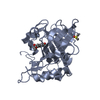
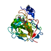
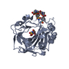
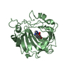
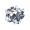
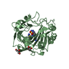
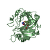
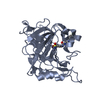
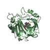
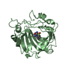
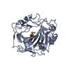
Structure of inhibitor binding to Carbonic Anhydrase I
Components
Carbonic anhydrase 1
Keywords
LYASE / zinc metalloenzyme
Function / homology
Function and homology information
hydro-lyase activity / Gene and protein expression by JAK-STAT signaling after Interleukin-12 stimulation / cyanamide hydratase / cyanamide hydratase activity / arylesterase activity / Reversible hydration of carbon dioxide / carbonic anhydrase / carbonate dehydratase activity / Erythrocytes take up oxygen and release carbon dioxide / Erythrocytes take up carbon dioxide and release oxygen ...hydro-lyase activity / Gene and protein expression by JAK-STAT signaling after Interleukin-12 stimulation / cyanamide hydratase / cyanamide hydratase activity / arylesterase activity / Reversible hydration of carbon dioxide / carbonic anhydrase / carbonate dehydratase activity / Erythrocytes take up oxygen and release carbon dioxide / Erythrocytes take up carbon dioxide and release oxygen / one-carbon metabolic process / extracellular exosome / zinc ion binding / cytosol Similarity search - Function
In the structure databanks used in Yorodumi, some data are registered as the other names, "COVID-19 virus" and "2019-nCoV". Here are the details of the virus and the list of structure data.
Jan 31, 2019. EMDB accession codes are about to change! (news from PDBe EMDB page)
EMDB accession codes are about to change! (news from PDBe EMDB page)
The allocation of 4 digits for EMDB accession codes will soon come to an end. Whilst these codes will remain in use, new EMDB accession codes will include an additional digit and will expand incrementally as the available range of codes is exhausted. The current 4-digit format prefixed with “EMD-” (i.e. EMD-XXXX) will advance to a 5-digit format (i.e. EMD-XXXXX), and so on. It is currently estimated that the 4-digit codes will be depleted around Spring 2019, at which point the 5-digit format will come into force.
The EM Navigator/Yorodumi systems omit the EMD- prefix.
Related info.:Q: What is EMD? / ID/Accession-code notation in Yorodumi/EM Navigator
Yorodumi is a browser for structure data from EMDB, PDB, SASBDB, etc.
This page is also the successor to EM Navigator detail page, and also detail information page/front-end page for Omokage search.
The word "yorodu" (or yorozu) is an old Japanese word meaning "ten thousand". "mi" (miru) is to see.
Related info.:EMDB / PDB / SASBDB / Comparison of 3 databanks / Yorodumi Search / Aug 31, 2016. New EM Navigator & Yorodumi / Yorodumi Papers / Jmol/JSmol / Function and homology information / Changes in new EM Navigator and Yorodumi
 Movie
Movie Controller


 Open data
 Basic information
 Components
 Keywords
 Function and homology information
 Authors
 Citation
 Structure visualization
 Downloads & links
 Other downloads
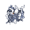


 PDBj
PDBj


 Assembly


 Mass: 65.409 Da / Num. of mol.: 2 / Source method: obtained synthetically / Formula: Zn
 Mass: 229.253 Da / Num. of mol.: 2 / Source method: obtained synthetically / Formula: C9H11NO4S
 Mass: 122.143 Da / Num. of mol.: 1 / Source method: obtained synthetically / Formula: C4H12NO3 / Comment: pH buffer*YM
 Mass: 22.990 Da / Num. of mol.: 1 / Source method: obtained synthetically / Formula: Na
 Sample preparation
 / Beamline: 5.0.2 / Wavelength: 1.4 Å
 Processing