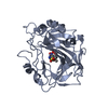
 Movie
Movie Controller

[English] 日本語
 Yorodumi
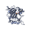
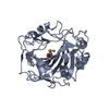
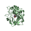
- PDB-2it4: X ray structure of the complex between Carbonic Anhydrase I and t... -
+ Open data

- Basic information
| Entry | Database: PDB / ID: 2it4 | ||||||
|---|---|---|---|---|---|---|---|
| Title | X ray structure of the complex between Carbonic Anhydrase I and the phosphonate antiviral drug foscarnet | ||||||
 Components Components | Carbonic anhydrase 1 | ||||||
 Keywords Keywords | LYASE / carbonic anhydrase / antiviral / crystal stucture | ||||||
| Function / homology |  Function and homology informationhydro-lyase activity / Gene and protein expression by JAK-STAT signaling after Interleukin-12 stimulation / cyanamide hydratase / cyanamide hydratase activity / arylesterase activity / Reversible hydration of carbon dioxide / carbonic anhydrase / carbonate dehydratase activity / Erythrocytes take up oxygen and release carbon dioxide / Erythrocytes take up carbon dioxide and release oxygen ...hydro-lyase activity / Gene and protein expression by JAK-STAT signaling after Interleukin-12 stimulation / cyanamide hydratase / cyanamide hydratase activity / arylesterase activity / Reversible hydration of carbon dioxide / carbonic anhydrase / carbonate dehydratase activity / Erythrocytes take up oxygen and release carbon dioxide / Erythrocytes take up carbon dioxide and release oxygen / one-carbon metabolic process / extracellular exosome / zinc ion binding / cytosol Function and homology informationhydro-lyase activity / Gene and protein expression by JAK-STAT signaling after Interleukin-12 stimulation / cyanamide hydratase / cyanamide hydratase activity / arylesterase activity / Reversible hydration of carbon dioxide / carbonic anhydrase / carbonate dehydratase activity / Erythrocytes take up oxygen and release carbon dioxide / Erythrocytes take up carbon dioxide and release oxygen ...hydro-lyase activity / Gene and protein expression by JAK-STAT signaling after Interleukin-12 stimulation / cyanamide hydratase / cyanamide hydratase activity / arylesterase activity / Reversible hydration of carbon dioxide / carbonic anhydrase / carbonate dehydratase activity / Erythrocytes take up oxygen and release carbon dioxide / Erythrocytes take up carbon dioxide and release oxygen / one-carbon metabolic process / extracellular exosome / zinc ion binding / cytosolSimilarity search - Function | ||||||
| Biological species |  Homo sapiens (human) Homo sapiens (human) | ||||||
| Method | X-RAY DIFFRACTION / MOLECULAR REPLACEMENT / Resolution: 2 Å | ||||||
 Authors Authors | Temperini, C. / Innocenti, A. / Guerri, A. / Scozzafava, A. / Supuran, C.T. | ||||||
 Citation Citation | Journal: Bioorg.Med.Chem.Lett. / Year: 2007 Title: Phosph(on)ate as a zinc-binding group in metalloenzyme inhibitors: X-ray crystal structure of the antiviral drug foscarnet complexed to human carbonic anhydrase I. Authors: Temperini, C. / Innocenti, A. / Guerri, A. / Scozzafava, A. / Rusconi, S. / Supuran, C.T. | ||||||
| History |
|
- Structure visualization
| Structure viewer | Molecule: MolmilJmol/JSmol |
|---|
- Downloads & links
-Download
| PDBx/mmCIF format | 2it4.cif.gz | 118.9 KB | Display | PDBx/mmCIF format |
|---|---|---|---|---|
| PDB format | pdb2it4.ent.gz | 91 KB | Display | PDB format |
| PDBx/mmJSON format | 2it4.json.gz | Tree view | PDBx/mmJSON format | |
| Others |  Other downloads Other downloads |
-Validation report
| Arichive directory | https://data.pdbj.org/pub/pdb/validation_reports/it/2it4ftp://data.pdbj.org/pub/pdb/validation_reports/it/2it4 | HTTPS FTP |
|---|
-Related structure data
| Related structure data | 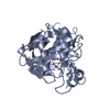 2fw4S S: Starting model for refinement |
|---|---|
| Similar structure data |


-Links
 PDBj
PDBj


- Assembly
| Deposited unit | 
| ||||||||
|---|---|---|---|---|---|---|---|---|---|
| 1 | 
| ||||||||
| 2 | 
| ||||||||
| Unit cell |
|
-Components
| #1: Protein | / Carbonic anhydrase I / Carbonate dehydratase I / CA-I Mass: 28404.629 Da / Num. of mol.: 2 / Source method: isolated from a natural source / Source: (natural) Homo sapiens (human) / References: UniProt: P00915, carbonic anhydrase#2: Chemical |   Mass: 65.409 Da / Num. of mol.: 2 / Source method: obtained synthetically / Formula: Zn Mass: 65.409 Da / Num. of mol.: 2 / Source method: obtained synthetically / Formula: Zn#3: Chemical | ChemComp-PPF / | Foscarnet  Mass: 126.005 Da / Num. of mol.: 1 / Source method: obtained synthetically / Formula: CH3O5P / Comment: medication, antivirus, inhibitor*YM Mass: 126.005 Da / Num. of mol.: 1 / Source method: obtained synthetically / Formula: CH3O5P / Comment: medication, antivirus, inhibitor*YM#4: Water | ChemComp-HOH / | Water Mass: 18.015 Da / Num. of mol.: 344 / Source method: isolated from a natural source / Formula: H2O Mass: 18.015 Da / Num. of mol.: 344 / Source method: isolated from a natural source / Formula: H2O |
|---|
-Experimental details
-Experiment
| Experiment | Method: X-RAY DIFFRACTION / Number of used crystals: 1 |
|---|
- Sample preparation
| Crystal | Density Matthews: 2.3 Å3/Da / Density % sol: 46.62 % |
|---|---|
| Crystal grow | Temperature: 295 K / Method: vapor diffusion, hanging drop / pH: 9 Details: 100mM Tris.HCl buffer pH 9.0, 25% (w/v) PEG 4000, LiCl4 0.4 M, 10% ethylene glycol , VAPOR DIFFUSION, HANGING DROP, temperature 22K |
-Data collection
| Diffraction | Mean temperature: 100 K |
|---|---|
| Diffraction source | Source: SEALED TUBE / Type: OXFORD DIFFRACTION ENHANCE ULTRA / Wavelength: 1.5418 Å |
| Detector | Type: OXFORD SAPPHIRE CCD / Detector: CCD / Date: Sep 13, 2006 |
| Radiation | Monochromator: capillary / Protocol: SINGLE WAVELENGTH / Monochromatic (M) / Laue (L): M / Scattering type: x-ray |
| Radiation wavelength | Wavelength: 1.5418 Å / Relative weight: 1 |
| Reflection | Resolution: 2→20 Å / Num. all: 211265 / Num. obs: 36020 / % possible obs: 97.1 % / Observed criterion σ(F): 2 / Observed criterion σ(I): 0 / Redundancy: 5.9 % / Rsym value: 0.127 / Net I/σ(I): 8.3 |
| Reflection shell | Resolution: 2→2.07 Å / Redundancy: 3.8 % / Mean I/σ(I) obs: 2.03 / Num. unique all: 3602 |
- Processing
| Software |
| ||||||||||||||||||
|---|---|---|---|---|---|---|---|---|---|---|---|---|---|---|---|---|---|---|---|
| Refinement | Method to determine structure: MOLECULAR REPLACEMENT Starting model: 2FW4 Resolution: 2→20 Å / Cross valid method: THROUGHOUT / σ(F): 0 / σ(I): 0
| ||||||||||||||||||
| Refinement step | Cycle: LAST / Resolution: 2→20 Å
| ||||||||||||||||||
| Refine LS restraints |
| ||||||||||||||||||
| LS refinement shell | Resolution: 2→2.07 Å
|
