 Movie
Movie Controller

[English] 日本語
 Yorodumi
- PDB-6g3v: Crystal structure of human carbonic anhydrase I in complex with t... -
+ Open data

- Basic information
| Entry | Database: PDB / ID: 6g3v | ||||||
|---|---|---|---|---|---|---|---|
| Title | Crystal structure of human carbonic anhydrase I in complex with the inhibitor famotidine | ||||||
 Components Components | Carbonic anhydrase 1 | ||||||
 Keywords Keywords | LYASE | ||||||
| Function / homology |  Function and homology informationhydro-lyase activity / Gene and protein expression by JAK-STAT signaling after Interleukin-12 stimulation / cyanamide hydratase / cyanamide hydratase activity / arylesterase activity / Reversible hydration of carbon dioxide / carbonic anhydrase / carbonate dehydratase activity / Erythrocytes take up oxygen and release carbon dioxide / Erythrocytes take up carbon dioxide and release oxygen ...hydro-lyase activity / Gene and protein expression by JAK-STAT signaling after Interleukin-12 stimulation / cyanamide hydratase / cyanamide hydratase activity / arylesterase activity / Reversible hydration of carbon dioxide / carbonic anhydrase / carbonate dehydratase activity / Erythrocytes take up oxygen and release carbon dioxide / Erythrocytes take up carbon dioxide and release oxygen / one-carbon metabolic process / extracellular exosome / zinc ion binding / cytosol Function and homology informationhydro-lyase activity / Gene and protein expression by JAK-STAT signaling after Interleukin-12 stimulation / cyanamide hydratase / cyanamide hydratase activity / arylesterase activity / Reversible hydration of carbon dioxide / carbonic anhydrase / carbonate dehydratase activity / Erythrocytes take up oxygen and release carbon dioxide / Erythrocytes take up carbon dioxide and release oxygen ...hydro-lyase activity / Gene and protein expression by JAK-STAT signaling after Interleukin-12 stimulation / cyanamide hydratase / cyanamide hydratase activity / arylesterase activity / Reversible hydration of carbon dioxide / carbonic anhydrase / carbonate dehydratase activity / Erythrocytes take up oxygen and release carbon dioxide / Erythrocytes take up carbon dioxide and release oxygen / one-carbon metabolic process / extracellular exosome / zinc ion binding / cytosolSimilarity search - Function | ||||||
| Biological species |  Homo sapiens (human) Homo sapiens (human) | ||||||
| Method | X-RAY DIFFRACTION / SYNCHROTRON / FOURIER SYNTHESIS / Resolution: 1.69 Å | ||||||
 Authors Authors | Ferraroni, M. / Supuran, C.T. / Angeli, A. | ||||||
 Citation Citation | Journal: ACS Med Chem Lett / Year: 2018 Title: Famotidine, an Antiulcer Agent, Strongly InhibitsHelicobacter pyloriand Human Carbonic Anhydrases. Authors: Angeli, A. / Ferraroni, M. / Supuran, C.T. | ||||||
| History |
|
- Structure visualization
| Structure viewer | Molecule: MolmilJmol/JSmol |
|---|
- Downloads & links
-Download
| PDBx/mmCIF format | 6g3v.cif.gz | 117.2 KB | Display | PDBx/mmCIF format |
|---|---|---|---|---|
| PDB format | pdb6g3v.ent.gz | 90 KB | Display | PDB format |
| PDBx/mmJSON format | 6g3v.json.gz | Tree view | PDBx/mmJSON format | |
| Others |  Other downloads Other downloads |
-Validation report
| Arichive directory | https://data.pdbj.org/pub/pdb/validation_reports/g3/6g3vftp://data.pdbj.org/pub/pdb/validation_reports/g3/6g3v | HTTPS FTP |
|---|
-Related structure data
| Related structure data |  6g3qC 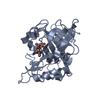 3lxeS S: Starting model for refinement C: citing same article ( |
|---|---|
| Similar structure data |


-Links
 PDBj
PDBj


- Assembly
| Deposited unit | 
| ||||||||
|---|---|---|---|---|---|---|---|---|---|
| 1 | 
| ||||||||
| 2 | 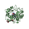
| ||||||||
| Unit cell |
|
-Components
| #1: Protein | / Carbonate dehydratase I / Carbonic anhydrase B / CAB / Carbonic anhydrase I / CA-I Mass: 28906.186 Da / Num. of mol.: 2 / Source method: isolated from a natural source / Details: erytrocytes / Source: (natural) Homo sapiens (human) / References: UniProt: P00915, carbonic anhydrase#2: Chemical |   Mass: 65.409 Da / Num. of mol.: 2 / Source method: obtained synthetically / Formula: Zn Mass: 65.409 Da / Num. of mol.: 2 / Source method: obtained synthetically / Formula: Zn#3: Chemical | Famotidine  Mass: 339.461 Da / Num. of mol.: 2 / Source method: obtained synthetically / Formula: C8H17N7O2S3 / Comment: medication, antagonist*YM Mass: 339.461 Da / Num. of mol.: 2 / Source method: obtained synthetically / Formula: C8H17N7O2S3 / Comment: medication, antagonist*YM#4: Chemical | Glycerol  Mass: 92.094 Da / Num. of mol.: 3 / Source method: obtained synthetically / Formula: C3H8O3 Mass: 92.094 Da / Num. of mol.: 3 / Source method: obtained synthetically / Formula: C3H8O3#5: Water | ChemComp-HOH / | Water Mass: 18.015 Da / Num. of mol.: 159 / Source method: isolated from a natural source / Formula: H2O Mass: 18.015 Da / Num. of mol.: 159 / Source method: isolated from a natural source / Formula: H2O |
|---|
-Experimental details
-Experiment
| Experiment | Method: X-RAY DIFFRACTION / Number of used crystals: 1 |
|---|
- Sample preparation
| Crystal | Density Matthews: 2.34 Å3/Da / Density % sol: 47.5 % |
|---|---|
| Crystal grow | Temperature: 296 K / Method: vapor diffusion / pH: 9 / Details: 28% PEG4000, 0.2 M sodium acetate, Tris 100 mM |
-Data collection
| Diffraction | Mean temperature: 100 K |
|---|---|
| Diffraction source | Source: SYNCHROTRON / Site: ESRF  / Beamline: ID23-2 / Wavelength: 0.873 Å / Beamline: ID23-2 / Wavelength: 0.873 Å |
| Detector | Type: DECTRIS PILATUS3 2M / Detector: PIXEL / Date: Jan 27, 2018 |
| Radiation | Protocol: SINGLE WAVELENGTH / Monochromatic (M) / Laue (L): M / Scattering type: x-ray |
| Radiation wavelength | Wavelength: 0.873 Å / Relative weight: 1 |
| Reflection | Resolution: 1.69→30 Å / Num. all: 158183 / Num. obs: 57352 / % possible obs: 93.5 % / Redundancy: 2.75 % / CC1/2: 0.99 / Rrim(I) all: 0.083 / Rsym value: 0.068 / Net I/σ(I): 8.3 |
| Reflection shell | Resolution: 1.69→1.8 Å / Redundancy: 2.2 % / Num. unique obs: 8041 / CC1/2: 0.37 / Rrim(I) all: 0.151 / Rsym value: 0.12 / % possible all: 82.6 |
- Processing
| Software |
| |||||||||||||||||||||||||||||||||||||||||||||
|---|---|---|---|---|---|---|---|---|---|---|---|---|---|---|---|---|---|---|---|---|---|---|---|---|---|---|---|---|---|---|---|---|---|---|---|---|---|---|---|---|---|---|---|---|---|---|
| Refinement | Method to determine structure: FOURIER SYNTHESIS Starting model: 3LXE Resolution: 1.69→27 Å / Cor.coef. Fo:Fc: 0.964 / Cor.coef. Fo:Fc free: 0.942 / SU B: 4.37 / SU ML: 0.128 / SU R Cruickshank DPI: 0.1275 / Cross valid method: THROUGHOUT / σ(F): 0 / ESU R: 0.128 / ESU R Free: 0.13 / Details: U VALUES : REFINED INDIVIDUALLY
| |||||||||||||||||||||||||||||||||||||||||||||
| Solvent computation | Ion probe radii: 0.8 Å / Shrinkage radii: 0.8 Å / VDW probe radii: 1.2 Å | |||||||||||||||||||||||||||||||||||||||||||||
| Displacement parameters | Biso max: 114.34 Å2 / Biso mean: 30.799 Å2 / Biso min: 14.93 Å2
| |||||||||||||||||||||||||||||||||||||||||||||
| Refinement step | Cycle: final / Resolution: 1.69→27 Å
| |||||||||||||||||||||||||||||||||||||||||||||
| Refine LS restraints |
| |||||||||||||||||||||||||||||||||||||||||||||
| LS refinement shell | Resolution: 1.693→1.737 Å / Rfactor Rfree error: 0 / Total num. of bins used: 20
|
