 Movie
Movie Controller

[English] 日本語
 Yorodumi
- PDB-1jv0: THE CRYSTAL STRUCTURE OF THE ZINC(II) ADDUCT OF THE CAI MICHIGAN ... -
+ Open data

- Basic information
| Entry | Database: PDB / ID: 1jv0 | ||||||
|---|---|---|---|---|---|---|---|
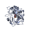
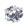
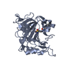
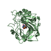
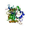
| Title | THE CRYSTAL STRUCTURE OF THE ZINC(II) ADDUCT OF THE CAI MICHIGAN 1 VARIANT | ||||||
 Components Components | CARBONIC ANHYDRASE I Carbonic anhydrase Carbonic anhydrase | ||||||
 Keywords Keywords | LYASE / 10-STRANDED-BETA-SHEET | ||||||
| Function / homology |  Function and homology informationhydro-lyase activity / Gene and protein expression by JAK-STAT signaling after Interleukin-12 stimulation / cyanamide hydratase / cyanamide hydratase activity / arylesterase activity / Reversible hydration of carbon dioxide / carbonic anhydrase / carbonate dehydratase activity / Erythrocytes take up oxygen and release carbon dioxide / Erythrocytes take up carbon dioxide and release oxygen ...hydro-lyase activity / Gene and protein expression by JAK-STAT signaling after Interleukin-12 stimulation / cyanamide hydratase / cyanamide hydratase activity / arylesterase activity / Reversible hydration of carbon dioxide / carbonic anhydrase / carbonate dehydratase activity / Erythrocytes take up oxygen and release carbon dioxide / Erythrocytes take up carbon dioxide and release oxygen / one-carbon metabolic process / extracellular exosome / zinc ion binding / cytosol Function and homology informationhydro-lyase activity / Gene and protein expression by JAK-STAT signaling after Interleukin-12 stimulation / cyanamide hydratase / cyanamide hydratase activity / arylesterase activity / Reversible hydration of carbon dioxide / carbonic anhydrase / carbonate dehydratase activity / Erythrocytes take up oxygen and release carbon dioxide / Erythrocytes take up carbon dioxide and release oxygen ...hydro-lyase activity / Gene and protein expression by JAK-STAT signaling after Interleukin-12 stimulation / cyanamide hydratase / cyanamide hydratase activity / arylesterase activity / Reversible hydration of carbon dioxide / carbonic anhydrase / carbonate dehydratase activity / Erythrocytes take up oxygen and release carbon dioxide / Erythrocytes take up carbon dioxide and release oxygen / one-carbon metabolic process / extracellular exosome / zinc ion binding / cytosolSimilarity search - Function | ||||||
| Biological species |  Homo sapiens (human) Homo sapiens (human) | ||||||
| Method | X-RAY DIFFRACTION / FOURIER SYNTHESIS / Resolution: 2 Å | ||||||
 Authors Authors | Briganti, F. / Ferraroni, M. / Chegwidden, W.R. / Scozzafava, A. / Supuran, C.T. / Tilli, S. | ||||||
 Citation Citation | Journal: Biochemistry / Year: 2002 Title: Crystal structure of a zinc-activated variant of human carbonic anhydrase I, CA I Michigan 1: evidence for a second zinc binding site involving arginine coordination Authors: Ferraroni, M. / Tilli, S. / Briganti, F. / Chegwidden, W.R. / Supuran, C.T. / Wiebauer, K.E. / Tashian, R.E. / Scozzafava, A. #1: Journal: Science / Year: 1962Title: New genetically determined molecular form of erythrocite esterase in man Authors: Shaw, C.R. / Syner, F.N. / Tashian, R.E. #2: Journal: HUM.MUTAT. / Year: 1994Title: Marked zinc activation of ester hydrolysis by a mutation, 67-HIS (CAT) To ARG (CGT), in the active site of human carbonic anhydrase I Authors: Chegwidden, W.R. / Wagner, L.E. / Venta, P.J. / Bergenhem, N.C. / Yu, Y.S. / Tashian, R.E. #3: Journal: GENE FAM.LSOZYME BULL. / Year: 1998Title: Point mutation in the variant of human carbonic anhydrase isozyme I (CAI MICHIGAN) active site decreases affinity for aromatic sulfonamide inhibitors Authors: Briganti, F. / Chegwidden, W.R. / Scozzafava, A. / Supuran, C.T. / Tashian, R.E. / Wiebauer, K.E. | ||||||
| History |
|
- Structure visualization
| Structure viewer | Molecule: MolmilJmol/JSmol |
|---|
- Downloads & links
-Download
| PDBx/mmCIF format | 1jv0.cif.gz | 126 KB | Display | PDBx/mmCIF format |
|---|---|---|---|---|
| PDB format | pdb1jv0.ent.gz | 95.4 KB | Display | PDB format |
| PDBx/mmJSON format | 1jv0.json.gz | Tree view | PDBx/mmJSON format | |
| Others |  Other downloads Other downloads |
-Validation report
| Arichive directory | https://data.pdbj.org/pub/pdb/validation_reports/jv/1jv0ftp://data.pdbj.org/pub/pdb/validation_reports/jv/1jv0 | HTTPS FTP |
|---|
-Related structure data
| Related structure data | 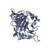 1j9wSC S: Starting model for refinement C: citing same article ( |
|---|---|
| Similar structure data |


-Links
 PDBj
PDBj


- Assembly
| Deposited unit | 
| ||||||||
|---|---|---|---|---|---|---|---|---|---|
| 1 | 
| ||||||||
| 2 | 
| ||||||||
| Unit cell |
| ||||||||
| Details | two molecules in the asymmetric units related by a translation |
-Components
| #1: Protein | Carbonic anhydrase / CA-I Mass: 28794.035 Da / Num. of mol.: 2 / Mutation: H67R Source method: isolated from a genetically manipulated source Source: (gene. exp.) Homo sapiens (human) / Plasmid: PKK232-2 / Production host:  Escherichia coli (E. coli) / Strain (production host): JM109 / References: UniProt: P00915, carbonic anhydrase Escherichia coli (E. coli) / Strain (production host): JM109 / References: UniProt: P00915, carbonic anhydrase#2: Chemical | ChemComp-ZN /   Mass: 65.409 Da / Num. of mol.: 6 / Source method: obtained synthetically / Formula: Zn Mass: 65.409 Da / Num. of mol.: 6 / Source method: obtained synthetically / Formula: Zn#3: Chemical | Chloride  Mass: 35.453 Da / Num. of mol.: 3 / Source method: obtained synthetically / Formula: Cl Mass: 35.453 Da / Num. of mol.: 3 / Source method: obtained synthetically / Formula: Cl#4: Chemical | Ethylene glycol  Mass: 62.068 Da / Num. of mol.: 2 / Source method: obtained synthetically / Formula: C2H6O2 Mass: 62.068 Da / Num. of mol.: 2 / Source method: obtained synthetically / Formula: C2H6O2#5: Water | ChemComp-HOH / | Water Mass: 18.015 Da / Num. of mol.: 440 / Source method: isolated from a natural source / Formula: H2O Mass: 18.015 Da / Num. of mol.: 440 / Source method: isolated from a natural source / Formula: H2O |
|---|
-Experimental details
-Experiment
| Experiment | Method: X-RAY DIFFRACTION / Number of used crystals: 1 |
|---|
- Sample preparation
| Crystal | Density Matthews: 2.25 Å3/Da / Density % sol: 45 % |
|---|---|
| Crystal grow | Temperature: 295 K / Method: vapor diffusion, hanging drop / pH: 9 Details: PEG 4000, LITIUM CHLORIDE, ETHYLENE GLICOL , pH 9.0, VAPOR DIFFUSION, HANGING DROP, temperature 295K |
-Data collection
| Diffraction | Mean temperature: 100 K |
|---|---|
| Diffraction source | Source: ROTATING ANODE / Type: MACSCIENCE M12XHF / Wavelength: 1.5418 Å |
| Detector | Type: BRUKER SMART 1000 / Detector: CCD / Date: Mar 21, 2001 / Details: GOBEL MIRRORS |
| Radiation | Monochromator: MIRRORS / Protocol: SINGLE WAVELENGTH / Monochromatic (M) / Laue (L): M / Scattering type: x-ray |
| Radiation wavelength | Wavelength: 1.5418 Å / Relative weight: 1 |
| Reflection | Resolution: 2→15 Å / Num. all: 35578 / Num. obs: 35578 / % possible obs: 94.9 % / Observed criterion σ(F): 0 / Observed criterion σ(I): 0 / Redundancy: 2.3 % / Biso Wilson estimate: 16.4 Å2 / Rsym value: 0.085 / Net I/σ(I): 8 |
| Reflection shell | Resolution: 2→2.071 Å / Mean I/σ(I) obs: 2.18 / Rsym value: 0.277 / % possible all: 81.9 |
- Processing
| Software |
| ||||||||||||||||||||||||||||||||||||||||||||||||
|---|---|---|---|---|---|---|---|---|---|---|---|---|---|---|---|---|---|---|---|---|---|---|---|---|---|---|---|---|---|---|---|---|---|---|---|---|---|---|---|---|---|---|---|---|---|---|---|---|---|
| Refinement | Method to determine structure: FOURIER SYNTHESIS Starting model: PDB ENTRY 1J9W Resolution: 2→15 Å / Isotropic thermal model: isotropic / Cross valid method: THROUGHOUT / σ(F): 0 / σ(I): 0 / Stereochemistry target values: Engh & Huber
| ||||||||||||||||||||||||||||||||||||||||||||||||
| Displacement parameters | Biso mean: 27.8 Å2 | ||||||||||||||||||||||||||||||||||||||||||||||||
| Refinement step | Cycle: LAST / Resolution: 2→15 Å
| ||||||||||||||||||||||||||||||||||||||||||||||||
| Refine LS restraints |
|
