 Movie
Movie Controller


+ Open data

- Basic information
| Entry | Database: PDB / ID: 8tob | ||||||
|---|---|---|---|---|---|---|---|
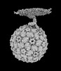
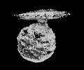
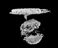
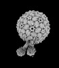
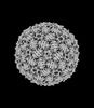
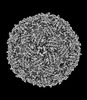
| Title | Acinetobacter GP16 Type IV pilus | ||||||
 Components Components | (Fimbrial protein) x 2 | ||||||
 Keywords Keywords |  CELL ADHESION / T4P / Competence CELL ADHESION / T4P / Competence | ||||||
| Function / homology |  Function and homology informationprotein secretion by the type II secretion system / type II protein secretion system complex / pilus / cell adhesion / membrane Function and homology informationprotein secretion by the type II secretion system / type II protein secretion system complex / pilus / cell adhesion / membraneSimilarity search - Function | ||||||
| Biological species |  Acinetobacter genomosp. 16BJ (bacteria) Acinetobacter genomosp. 16BJ (bacteria) | ||||||
| Method | ELECTRON MICROSCOPY / helical reconstruction / cryo EM / Resolution: 3.14 Å | ||||||
 Authors Authors | Meng, R. / Xing, Z. / Zhang, J. | ||||||
| Funding support |  United States, 1items United States, 1items
| ||||||
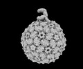
 Citation Citation | Journal: Nat Commun / Year: 2024 Title: Structural basis of Acinetobacter type IV pili targeting by an RNA virus. Authors: Ran Meng / Zhongliang Xing / Jeng-Yih Chang / Zihao Yu / Jirapat Thongchol / Wen Xiao / Yuhang Wang / Karthik Chamakura / Zhiqi Zeng / Fengbin Wang / Ry Young / Lanying Zeng / Junjie Zhang / Abstract: Acinetobacters pose a significant threat to human health, especially those with weakened immune systems. Type IV pili of acinetobacters play crucial roles in virulence and antibiotic resistance. ...Acinetobacters pose a significant threat to human health, especially those with weakened immune systems. Type IV pili of acinetobacters play crucial roles in virulence and antibiotic resistance. Single-stranded RNA bacteriophages target the bacterial retractile pili, including type IV. Our study delves into the interaction between Acinetobacter phage AP205 and type IV pili. Using cryo-electron microscopy, we solve structures of the AP205 virion with an asymmetric dimer of maturation proteins, the native Acinetobacter type IV pili bearing a distinct post-translational pilin cleavage, and the pili-bound AP205 showing its maturation proteins adapted to pilin modifications, allowing each phage to bind to one or two pili. Leveraging these results, we develop a 20-kilodalton AP205-derived protein scaffold targeting type IV pili in situ, with potential for research and diagnostics. | ||||||
| History |
|
- Structure visualization
| Structure viewer | Molecule: MolmilJmol/JSmol |
|---|
- Downloads & links
-Download
| PDBx/mmCIF format | 8tob.cif.gz | 472 KB | Display | PDBx/mmCIF format |
|---|---|---|---|---|
| PDB format | pdb8tob.ent.gz | Display | PDB format | |
| PDBx/mmJSON format | 8tob.json.gz | Tree view | PDBx/mmJSON format | |
| Others |  Other downloads Other downloads |
-Validation report
| Arichive directory | https://data.pdbj.org/pub/pdb/validation_reports/to/8tobftp://data.pdbj.org/pub/pdb/validation_reports/to/8tob | HTTPS FTP |
|---|
-Related structure data
| Related structure data | 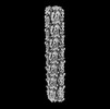 41442MC  8tocC 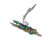 8tv9C 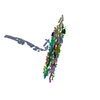 8tvaC  8tw2C  8twcC M: map data used to model this data C: citing same article ( |
|---|---|
| Similar structure data |
-Links
 PDBj
PDBj
- Assembly
| Deposited unit | 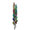
|
|---|---|
| 1 |
|
-Components
| #1: Protein | Mass: 7470.747 Da / Num. of mol.: 22 / Fragment: residues 9-78 / Source method: isolated from a natural source / Source: (natural) Acinetobacter genomosp. 16BJ (bacteria) / References: UniProt: N9RQW9#2: Protein | Mass: 6999.778 Da / Num. of mol.: 22 / Fragment: residues 79-147 / Source method: isolated from a natural source / Source: (natural) Acinetobacter genomosp. 16BJ (bacteria) / References: UniProt: N9RQW9Has ligand of interest | N | |
|---|
-Experimental details
-Experiment
| Experiment | Method: ELECTRON MICROSCOPY |
|---|---|
| EM experiment | Aggregation state: FILAMENT / 3D reconstruction method: helical reconstruction |
- Sample preparation
| Component | Name: Acinetobacter genomosp.16 Tyoe IV pilus / Type: COMPLEX / Details: Filamentous Type IV pilus / Entity ID: all / Source: NATURAL | |||||||||||||||
|---|---|---|---|---|---|---|---|---|---|---|---|---|---|---|---|---|
| Molecular weight | Value: 11 kDa/nm / Experimental value: NO | |||||||||||||||
| Source (natural) | Organism: Acinetobacter genomosp. 16BJ (bacteria) | |||||||||||||||
| Buffer solution | pH: 8 / Details: 20mM Tris-HCl, 200mM NaCl, pH 8.0 | |||||||||||||||
| Buffer component |
| |||||||||||||||
| Specimen | Conc.: 1.35 mg/ml / Embedding applied: NO / Shadowing applied: NO / Staining applied: NO / Vitrification applied: YES / Details: vitreous Typy IV pilus | |||||||||||||||
| Specimen support | Grid material: COPPER / Grid mesh size: 300 divisions/in. / Grid type: EMS Lacey Carbon | |||||||||||||||
| Vitrification | Instrument: FEI VITROBOT MARK III / Cryogen name: ETHANE / Humidity: 100 % / Chamber temperature: 277 K / Details: 3uL sample applied to a QuantiFoil R2/1 grid |
- Electron microscopy imaging
| Experimental equipment |  Model: Titan Krios / Image courtesy: FEI Company |
|---|---|
| Microscopy | Model: FEI TITAN KRIOS |
| Electron gun | Electron source: FIELD EMISSION GUN / Accelerating voltage: 300 kV / Illumination mode: FLOOD BEAM |
| Electron lens | Mode: BRIGHT FIELDBright-field microscopy / Nominal magnification: 135000 X / Nominal defocus max: 2000 nm / Nominal defocus min: 200 nm / Cs: 2.7 mm |
| Specimen holder | Cryogen: NITROGEN |
| Image recording | Electron dose: 50 e/Å2 / Film or detector model: GATAN K3 BIOQUANTUM (6k x 4k) |
- Processing
| EM software | Name: PHENIX / Version: 1.19.2_4158: / Category: model refinement | ||||||||||||||||||||||||
|---|---|---|---|---|---|---|---|---|---|---|---|---|---|---|---|---|---|---|---|---|---|---|---|---|---|
| CTF correction | Type: NONE | ||||||||||||||||||||||||
| Helical symmerty | Angular rotation/subunit: 92 ° / Axial rise/subunit: 10 Å / Axial symmetry: C1 | ||||||||||||||||||||||||
| 3D reconstruction | Resolution: 3.14 Å / Resolution method: FSC 0.143 CUT-OFF / Num. of particles: 900000 / Symmetry type: HELICAL | ||||||||||||||||||||||||
| Atomic model building | Protocol: BACKBONE TRACE | ||||||||||||||||||||||||
| Refine LS restraints |
|
