Movie
Movie Controller

[English] 日本語
 Yorodumi
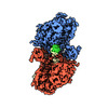
- PDB-9bh9: Human DNA polymerase theta helicase domain dimer bound to DNA in ... -
+ Open data

- Basic information
| Entry | Database: PDB / ID: 9bh9 | |||||||||
|---|---|---|---|---|---|---|---|---|---|---|
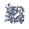
| Title | Human DNA polymerase theta helicase domain dimer bound to DNA in the microhomology aligning conformation | |||||||||
 Components Components |
| |||||||||
 Keywords Keywords |  DNA BINDING PROTEIN / DNA repair / TMEJ / MMEJ DNA BINDING PROTEIN / DNA repair / TMEJ / MMEJ | |||||||||
| Function / homology |  Function and homology information Function and homology informationsingle-stranded DNA endodeoxyribonuclease activity / double-strand break repair via alternative nonhomologous end joining / HDR through MMEJ (alt-NHEJ) / single-stranded DNA helicase activity / negative regulation of double-strand break repair via homologous recombination / replication fork processing / site of DNA damage / 5'-deoxyribose-5-phosphate lyase activity / somatic hypermutation of immunoglobulin genes / error-prone translesion synthesis ...single-stranded DNA endodeoxyribonuclease activity / double-strand break repair via alternative nonhomologous end joining / HDR through MMEJ (alt-NHEJ) / single-stranded DNA helicase activity / negative regulation of double-strand break repair via homologous recombination / replication fork processing / site of DNA damage / 5'-deoxyribose-5-phosphate lyase activity / somatic hypermutation of immunoglobulin genes / error-prone translesion synthesis / DNA helicase activity / base-excision repair / protein homooligomerization / RNA-directed DNA polymerase / RNA-directed DNA polymerase activity / double-strand break repair / site of double-strand break / DNA helicase / damaged DNA binding / DNA-directed DNA polymerase / DNA-directed DNA polymerase activity / DNA repair / DNA damage response / chromatin binding / Golgi apparatus / magnesium ion binding / ATP hydrolysis activity / nucleoplasm / ATP binding / identical protein binding / cytosolSimilarity search - Function | |||||||||
| Biological species |  Homo sapiens (human) Homo sapiens (human)synthetic construct (others) | |||||||||
| Method | ELECTRON MICROSCOPY / single particle reconstruction / cryo EM / Resolution: 3.5 Å | |||||||||
 Authors Authors | Zerio, C.J. / Lander, G.C. | |||||||||
| Funding support |  United States, 2items United States, 2items
| |||||||||
 Citation Citation | Journal: To Be Published Title: Human polymerase theta helicase positions DNA microhomologies for double-strand break repair Authors: Zerio, C.J. / Bai, Y. / Sosa-Alvarado, B.A. / Guzi, T. / Lander, G.C. | |||||||||
| History |
|
- Structure visualization
| Structure viewer | Molecule: MolmilJmol/JSmol |
|---|
- Downloads & links
-Download
| PDBx/mmCIF format | 9bh9.cif.gz | 302.2 KB | Display | PDBx/mmCIF format |
|---|---|---|---|---|
| PDB format | pdb9bh9.ent.gz | 237.7 KB | Display | PDB format |
| PDBx/mmJSON format | 9bh9.json.gz | Tree view | PDBx/mmJSON format | |
| Others |  Other downloads Other downloads |
-Validation report
| Arichive directory | https://data.pdbj.org/pub/pdb/validation_reports/bh/9bh9ftp://data.pdbj.org/pub/pdb/validation_reports/bh/9bh9 | HTTPS FTP |
|---|
-Related structure data
| Related structure data |  44537MC  9bh6C 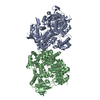 9bh7C 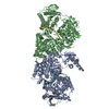 9bh8C 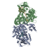 9bhaC M: map data used to model this data C: citing same article ( |
|---|---|
| Similar structure data |


-Links
 PDBj
PDBj


- Assembly
| Deposited unit | 
|
|---|---|
| 1 |
|
-Components
| #1: Protein | POLQ / DNA polymerase eta Mass: 99671.344 Da / Num. of mol.: 2 Source method: isolated from a genetically manipulated source Source: (gene. exp.) Homo sapiens (human) / Gene: POLQ, POLH / Plasmid: pET-Duet-1 / Production host:  Escherichia coli BL21(DE3) (bacteria) / Variant (production host): Rosetta Escherichia coli BL21(DE3) (bacteria) / Variant (production host): RosettaReferences: UniProt: O75417, DNA helicase, DNA-directed DNA polymerase, RNA-directed DNA polymerase#2: DNA chain | Mass: 17264.012 Da / Num. of mol.: 2 / Source method: obtained synthetically / Source: (synth.) synthetic construct (others) |
|---|
-Experimental details
-Experiment
| Experiment | Method: ELECTRON MICROSCOPY |
|---|---|
| EM experiment | Aggregation state: PARTICLE / 3D reconstruction method: single particle reconstruction |
- Sample preparation
| Component |
| ||||||||||||||||||||||||
|---|---|---|---|---|---|---|---|---|---|---|---|---|---|---|---|---|---|---|---|---|---|---|---|---|---|
| Molecular weight |
| ||||||||||||||||||||||||
| Source (natural) |
| ||||||||||||||||||||||||
| Source (recombinant) |
| ||||||||||||||||||||||||
| Buffer solution | pH: 7.5 | ||||||||||||||||||||||||
| Buffer component |
| ||||||||||||||||||||||||
| Specimen | Conc.: 1.25 mg/ml / Embedding applied: NO / Shadowing applied: NO / Staining applied: NO / Vitrification applied: YES Details: We added 2X pre-annealed DNA to the protein, lowered the NaCl concentration to 100 mM, and purified the DNA-bound complex by SEC. | ||||||||||||||||||||||||
| Specimen support | Details: Grids were glow discharged under vacuum for 30 s at 15 mA in a Pelco easiGlow 91000 Glow Discharge Cleaning System. Grid material: COPPER / Grid mesh size: 300 divisions/in. / Grid type: Quantifoil R1.2/1.3 | ||||||||||||||||||||||||
| Vitrification | Instrument: HOMEMADE PLUNGER / Cryogen name: ETHANE / Humidity: 95 % / Chamber temperature: 277 K Details: 3 microliters of sample was applied to the surface of the grid, blotted for 2 seconds after the liquid spot on the filter paper stopped spreading, and the grid was plunged into a liquid ...Details: 3 microliters of sample was applied to the surface of the grid, blotted for 2 seconds after the liquid spot on the filter paper stopped spreading, and the grid was plunged into a liquid ethane bath cooled by liquid nitrogen. |
- Electron microscopy imaging
| Experimental equipment |  Model: Titan Krios / Image courtesy: FEI Company |
|---|---|
| Microscopy | Model: FEI TITAN KRIOS |
| Electron gun | Electron source: FIELD EMISSION GUN / Accelerating voltage: 300 kV / Illumination mode: FLOOD BEAM |
| Electron lens | Mode: BRIGHT FIELDBright-field microscopy / Nominal magnification: 105000 X / Calibrated magnification: 60024 X / Nominal defocus max: 1000 nm / Nominal defocus min: 1000 nm / Calibrated defocus min: 300 nm / Calibrated defocus max: 2500 nm / Cs: 2.7 mm / C2 aperture diameter: 150 µm / Alignment procedure: COMA FREE |
| Specimen holder | Cryogen: NITROGEN / Specimen holder model: FEI TITAN KRIOS AUTOGRID HOLDER / Temperature (max): 77 K / Temperature (min): 70 K |
| Image recording | Average exposure time: 1.4 sec. / Electron dose: 50 e/Å2 / Film or detector model: GATAN K3 BIOQUANTUM (6k x 4k) / Num. of grids imaged: 1 / Num. of real images: 12440 |
| EM imaging optics | Energyfilter slit width: 20 eV |
| Image scans | Sampling size: 5 µm / Width: 5760 / Height: 4092 |
- Processing
| EM software |
| |||||||||||||||||||||||||||||||||||||||||||||||||||||||
|---|---|---|---|---|---|---|---|---|---|---|---|---|---|---|---|---|---|---|---|---|---|---|---|---|---|---|---|---|---|---|---|---|---|---|---|---|---|---|---|---|---|---|---|---|---|---|---|---|---|---|---|---|---|---|---|---|
| CTF correction | Type: PHASE FLIPPING AND AMPLITUDE CORRECTION | |||||||||||||||||||||||||||||||||||||||||||||||||||||||
| Particle selection | Num. of particles selected: 5424691 | |||||||||||||||||||||||||||||||||||||||||||||||||||||||
| Symmetry | Point symmetry: C1 (asymmetric) | |||||||||||||||||||||||||||||||||||||||||||||||||||||||
| 3D reconstruction | Resolution: 3.5 Å / Resolution method: FSC 0.143 CUT-OFF / Num. of particles: 107353 / Algorithm: FOURIER SPACE Details: Non-uniform refinement and local filtering by resolution Symmetry type: POINT | |||||||||||||||||||||||||||||||||||||||||||||||||||||||
| Atomic model building | B value: 67.19 / Protocol: FLEXIBLE FIT / Space: REAL / Target criteria: Cross-correlation coefficient Details: We modeled DNA bases into sharpened density when possible. We modeled the DNA backbone into locally filtered density in low resolution areas. | |||||||||||||||||||||||||||||||||||||||||||||||||||||||
| Atomic model building | 3D fitting-ID: 1 / Source name: PDB / Type: experimental model
|