Movie
Movie Controller

[English] 日本語
 Yorodumi
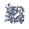
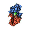
- PDB-9bh6: Human DNA polymerase theta helicase domain tetramer in the apo form -
+ Open data

- Basic information
| Entry | Database: PDB / ID: 9bh6 | |||||||||
|---|---|---|---|---|---|---|---|---|---|---|
| Title | Human DNA polymerase theta helicase domain tetramer in the apo form | |||||||||
 Components Components | DNA polymerase theta POLQ POLQ | |||||||||
 Keywords Keywords | DNA BINDING PROTEIN / DNA repair / TMEJ / MMEJ | |||||||||
| Function / homology |  Function and homology information Function and homology informationsingle-stranded DNA endodeoxyribonuclease activity / double-strand break repair via alternative nonhomologous end joining / HDR through MMEJ (alt-NHEJ) / single-stranded DNA helicase activity / negative regulation of double-strand break repair via homologous recombination / replication fork processing / site of DNA damage / 5'-deoxyribose-5-phosphate lyase activity / somatic hypermutation of immunoglobulin genes / error-prone translesion synthesis ...single-stranded DNA endodeoxyribonuclease activity / double-strand break repair via alternative nonhomologous end joining / HDR through MMEJ (alt-NHEJ) / single-stranded DNA helicase activity / negative regulation of double-strand break repair via homologous recombination / replication fork processing / site of DNA damage / 5'-deoxyribose-5-phosphate lyase activity / somatic hypermutation of immunoglobulin genes / error-prone translesion synthesis / DNA helicase activity / base-excision repair / protein homooligomerization / RNA-directed DNA polymerase / RNA-directed DNA polymerase activity / double-strand break repair / site of double-strand break / DNA helicase / damaged DNA binding / DNA-directed DNA polymerase / DNA-directed DNA polymerase activity / DNA repair / DNA damage response / chromatin binding / Golgi apparatus / magnesium ion binding / ATP hydrolysis activity / nucleoplasm / ATP binding / identical protein binding / cytosolSimilarity search - Function | |||||||||
| Biological species |  Homo sapiens (human) Homo sapiens (human) | |||||||||
| Method | ELECTRON MICROSCOPY / single particle reconstruction / cryo EM / Resolution: 3.3 Å | |||||||||
 Authors Authors | Zerio, C.J. / Lander, G.C. | |||||||||
| Funding support |  United States, 2items United States, 2items
| |||||||||
 Citation Citation | Journal: To Be Published Title: Human polymerase theta helicase positions DNA microhomologies for double-strand break repair Authors: Zerio, C.J. / Bai, Y. / Sosa-Alvarado, B.A. / Guzi, T. / Lander, G.C. | |||||||||
| History |
|
- Structure visualization
| Structure viewer | Molecule: MolmilJmol/JSmol |
|---|
- Downloads & links
-Download
| PDBx/mmCIF format | 9bh6.cif.gz | 598.3 KB | Display | PDBx/mmCIF format |
|---|---|---|---|---|
| PDB format | pdb9bh6.ent.gz | 486.1 KB | Display | PDB format |
| PDBx/mmJSON format | 9bh6.json.gz | Tree view | PDBx/mmJSON format | |
| Others |  Other downloads Other downloads |
-Validation report
| Arichive directory | https://data.pdbj.org/pub/pdb/validation_reports/bh/9bh6ftp://data.pdbj.org/pub/pdb/validation_reports/bh/9bh6 | HTTPS FTP |
|---|
-Related structure data
| Related structure data |  44534MC 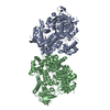 9bh7C 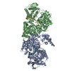 9bh8C 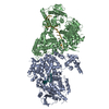 9bh9C 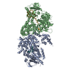 9bhaC M: map data used to model this data C: citing same article ( |
|---|---|
| Similar structure data |


-Links
 PDBj
PDBj


- Assembly
| Deposited unit | 
|
|---|---|
| 1 |
|
-Components
| #1: Protein | POLQ / DNA polymerase eta Mass: 99671.344 Da / Num. of mol.: 4 Source method: isolated from a genetically manipulated source Source: (gene. exp.) Homo sapiens (human) / Gene: POLQ, POLH / Plasmid: pET-Duet-1 / Production host:  Escherichia coli BL21(DE3) (bacteria) / Variant (production host): Rosetta Escherichia coli BL21(DE3) (bacteria) / Variant (production host): RosettaReferences: UniProt: O75417, DNA helicase, DNA-directed DNA polymerase, RNA-directed DNA polymerase |
|---|
-Experimental details
-Experiment
| Experiment | Method: ELECTRON MICROSCOPY |
|---|---|
| EM experiment | Aggregation state: PARTICLE / 3D reconstruction method: single particle reconstruction |
- Sample preparation
| Component | Name: Human DNA polymerase theta helicase domain / Type: COMPLEX / Entity ID: all / Source: RECOMBINANT | ||||||||||||||||||||
|---|---|---|---|---|---|---|---|---|---|---|---|---|---|---|---|---|---|---|---|---|---|
| Molecular weight | Value: 0.4 MDa / Experimental value: NO | ||||||||||||||||||||
| Source (natural) | Organism: Homo sapiens (human) | ||||||||||||||||||||
| Source (recombinant) | Organism: Escherichia coli (E. coli) / Strain: Rosetta(DE3) / Plasmid: pET-Duet-1 | ||||||||||||||||||||
| Buffer solution | pH: 7.5 | ||||||||||||||||||||
| Buffer component |
| ||||||||||||||||||||
| Specimen | Conc.: 0.9 mg/ml / Embedding applied: NO / Shadowing applied: NO / Staining applied: NO / Vitrification applied: YES | ||||||||||||||||||||
| Specimen support | Details: Grids were glow discharged under vacuum for 30 s at 15 mA in a Pelco easiGlow 91000 Glow Discharge Cleaning System. Grid material: COPPER / Grid mesh size: 300 divisions/in. / Grid type: Quantifoil R1.2/1.3 | ||||||||||||||||||||
| Vitrification | Instrument: HOMEMADE PLUNGER / Cryogen name: ETHANE / Humidity: 95 % / Chamber temperature: 277 K Details: 3 microliters of sample was applied to the surface of the grid, blotted with Whatman 1 filter paper until 2 seconds after the liquid spot on the filter paper stopped spreading, and the grid ...Details: 3 microliters of sample was applied to the surface of the grid, blotted with Whatman 1 filter paper until 2 seconds after the liquid spot on the filter paper stopped spreading, and the grid was plunged into a liquid ethane pool cooled by liquid nitrogen using a manual plunge freezer. |
- Electron microscopy imaging
| Experimental equipment |  Model: Titan Krios / Image courtesy: FEI Company |
|---|---|
| Microscopy | Model: FEI TITAN KRIOS |
| Electron gun | Electron source: FIELD EMISSION GUN / Accelerating voltage: 300 kV / Illumination mode: FLOOD BEAM |
| Electron lens | Mode: BRIGHT FIELDBright-field microscopy / Nominal magnification: 105000 X / Calibrated magnification: 60024 X / Nominal defocus max: 1000 nm / Nominal defocus min: 1000 nm / Calibrated defocus min: 300 nm / Calibrated defocus max: 2000 nm / Cs: 2.7 mm / C2 aperture diameter: 150 µm / Alignment procedure: COMA FREE |
| Specimen holder | Cryogen: NITROGEN / Specimen holder model: FEI TITAN KRIOS AUTOGRID HOLDER / Temperature (max): 77 K / Temperature (min): 70 K |
| Image recording | Average exposure time: 0.9 sec. / Electron dose: 50 e/Å2 / Film or detector model: GATAN K3 BIOQUANTUM (6k x 4k) / Num. of grids imaged: 1 / Num. of real images: 3906 |
| Image scans | Sampling size: 5 µm / Width: 5760 / Height: 4092 |
- Processing
| EM software |
| |||||||||||||||||||||||||||||||||||||||||||||||||||||||
|---|---|---|---|---|---|---|---|---|---|---|---|---|---|---|---|---|---|---|---|---|---|---|---|---|---|---|---|---|---|---|---|---|---|---|---|---|---|---|---|---|---|---|---|---|---|---|---|---|---|---|---|---|---|---|---|---|
| CTF correction | Type: PHASE FLIPPING AND AMPLITUDE CORRECTION | |||||||||||||||||||||||||||||||||||||||||||||||||||||||
| Particle selection | Num. of particles selected: 1474207 | |||||||||||||||||||||||||||||||||||||||||||||||||||||||
| Symmetry | Point symmetry: D2 (2x2 fold dihedral) | |||||||||||||||||||||||||||||||||||||||||||||||||||||||
| 3D reconstruction | Resolution: 3.3 Å / Resolution method: FSC 0.143 CUT-OFF / Num. of particles: 208545 / Algorithm: FOURIER SPACE Details: We performed local refinement of a single protomer and then multiplied the output protomer map to fit the D2 symmetric tetramer map. Symmetry type: POINT | |||||||||||||||||||||||||||||||||||||||||||||||||||||||
| Atomic model building | B value: 54.43 / Protocol: FLEXIBLE FIT / Space: REAL / Target criteria: Cross-correlation coefficient Details: We used ISOLDE to flexibly fit one protomer into the locally refined protomer map. The output protomer model was multiplied and we used ISOLDE to fit four protomers into the tetramer map. | |||||||||||||||||||||||||||||||||||||||||||||||||||||||
| Atomic model building | PDB-ID: 5A9J Pdb chain-ID: A / Accession code: 5A9J / Chain residue range: 67-892 / Pdb chain residue range: 67-892 / Source name: PDB / Type: experimental model |