Movie
Movie Controller


+ Open data

- Basic information
| Entry | Database: PDB / ID: 8pqv | ||||||||||||
|---|---|---|---|---|---|---|---|---|---|---|---|---|---|
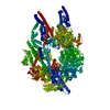
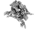
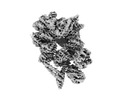
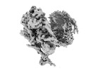
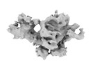
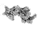
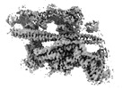
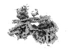
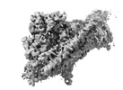
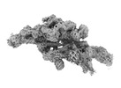
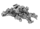
| Title | Cytoplasmic dynein-1 motor domain in post-powerstroke state | ||||||||||||
 Components Components | Cytoplasmic dynein 1 heavy chain 1 | ||||||||||||
 Keywords Keywords |  MOTOR PROTEIN / Dynein / AAA-Atpase MOTOR PROTEIN / Dynein / AAA-Atpase | ||||||||||||
| Function / homology |  Function and homology information Function and homology informationpositive regulation of intracellular transport / regulation of metaphase plate congression / establishment of spindle localization / positive regulation of spindle assembly / P-body assembly / dynein complex / COPI-independent Golgi-to-ER retrograde traffic / minus-end-directed microtubule motor activity / cytoplasmic dynein complex / retrograde axonal transport ...positive regulation of intracellular transport / regulation of metaphase plate congression / establishment of spindle localization / positive regulation of spindle assembly / P-body assembly / dynein complex / COPI-independent Golgi-to-ER retrograde traffic / minus-end-directed microtubule motor activity / cytoplasmic dynein complex / retrograde axonal transport / dynein light intermediate chain binding / nuclear migration / dynein intermediate chain binding / cytoplasmic microtubule / Amplification of signal from unattached kinetochores via a MAD2 inhibitory signal / COPI-mediated anterograde transport / stress granule assembly / cytoplasmic microtubule organization / Mitotic Prometaphase / regulation of mitotic spindle organization / EML4 and NUDC in mitotic spindle formation / axon cytoplasm / Loss of Nlp from mitotic centrosomes / Loss of proteins required for interphase microtubule organization from the centrosome / Recruitment of mitotic centrosome proteins and complexes / Resolution of Sister Chromatid Cohesion / Recruitment of NuMA to mitotic centrosomes / HSP90 chaperone cycle for steroid hormone receptors (SHR) in the presence of ligand / Anchoring of the basal body to the plasma membrane / MHC class II antigen presentation / AURKA Activation by TPX2 / mitotic spindle organization / filopodium / RHO GTPases Activate Formins / Aggrephagy / HCMV Early Events / Separation of Sister Chromatids / azurophil granule lumen / Regulation of PLK1 Activity at G2/M Transition / positive regulation of cold-induced thermogenesis / cell cortex / microtubule / cell division / centrosome / Neutrophil degranulation / ATP hydrolysis activity / RNA binding / extracellular exosome / extracellular region / ATP binding / membrane / cytosolSimilarity search - Function | ||||||||||||
| Biological species |  Homo sapiens (human) Homo sapiens (human) | ||||||||||||
| Method | ELECTRON MICROSCOPY / single particle reconstruction / cryo EM / Resolution: 4 Å | ||||||||||||
 Authors Authors | Singh, K. / Lau, C.K. / Manigrasso, G. / Gassmann, R. / Carter, A.P. | ||||||||||||
| Funding support |  United Kingdom, European Union, 3items United Kingdom, European Union, 3items
| ||||||||||||
 Citation Citation | Journal: Science / Year: 2024 Title: Molecular mechanism of dynein-dynactin complex assembly by LIS1. Authors: Kashish Singh / Clinton K Lau / Giulia Manigrasso / José B Gama / Reto Gassmann / Andrew P Carter /  Abstract: Cytoplasmic dynein is a microtubule motor vital for cellular organization and division. It functions as a ~4-megadalton complex containing its cofactor dynactin and a cargo-specific coiled-coil ...Cytoplasmic dynein is a microtubule motor vital for cellular organization and division. It functions as a ~4-megadalton complex containing its cofactor dynactin and a cargo-specific coiled-coil adaptor. However, how dynein and dynactin recognize diverse adaptors, how they interact with each other during complex formation, and the role of critical regulators such as lissencephaly-1 (LIS1) protein (LIS1) remain unclear. In this study, we determined the cryo-electron microscopy structure of dynein-dynactin on microtubules with LIS1 and the lysosomal adaptor JIP3. This structure reveals the molecular basis of interactions occurring during dynein activation. We show how JIP3 activates dynein despite its atypical architecture. Unexpectedly, LIS1 binds dynactin's p150 subunit, tethering it along the length of dynein. Our data suggest that LIS1 and p150 constrain dynein-dynactin to ensure efficient complex formation. | ||||||||||||
| History |
|
- Structure visualization
| Structure viewer | Molecule: MolmilJmol/JSmol |
|---|
- Downloads & links
-Download
| PDBx/mmCIF format | 8pqv.cif.gz | 564.6 KB | Display | PDBx/mmCIF format |
|---|---|---|---|---|
| PDB format | pdb8pqv.ent.gz | 439.3 KB | Display | PDB format |
| PDBx/mmJSON format | 8pqv.json.gz | Tree view | PDBx/mmJSON format | |
| Others |  Other downloads Other downloads |
-Validation report
| Arichive directory | https://data.pdbj.org/pub/pdb/validation_reports/pq/8pqvftp://data.pdbj.org/pub/pdb/validation_reports/pq/8pqv | HTTPS FTP |
|---|
-Related structure data
| Related structure data |  17825MC  8pqwC  8pqyC 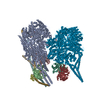 8pqzC  8pr0C  8pr1C  8pr2C  8pr3C 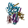 8pr4C 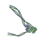 8pr5C  8ptkC M: map data used to model this data C: citing same article ( |
|---|---|
| Similar structure data |
-Links
 PDBj
PDBj


- Assembly
| Deposited unit | 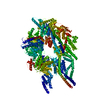
|
|---|---|
| 1 |
|
-Components
| #1: Protein | Mass: 533055.125 Da / Num. of mol.: 1 / Mutation: R1567E, K1610E Source method: isolated from a genetically manipulated source Source: (gene. exp.) Homo sapiens (human) / Gene: DYNC1H1, DHC1, DNCH1, DNCL, DNECL, DYHC, KIAA0325 / Production host:   Spodoptera frugiperda (fall armyworm) / References: UniProt: Q14204 Spodoptera frugiperda (fall armyworm) / References: UniProt: Q14204 |
|---|---|
| Has ligand of interest | Y |
-Experimental details
-Experiment
| Experiment | Method: ELECTRON MICROSCOPY |
|---|---|
| EM experiment | Aggregation state: PARTICLE / 3D reconstruction method: single particle reconstruction |
- Sample preparation
| Component | Name: Cytoplasmic dynein-1Dynein / Type: COMPLEX / Entity ID: all / Source: RECOMBINANT |
|---|---|
| Source (natural) | Organism: Homo sapiens (human) |
| Source (recombinant) | Organism: Spodoptera frugiperda (fall armyworm) |
| Buffer solution | pH: 7.2 |
| Specimen | Embedding applied: NO / Shadowing applied: NO / Staining applied: NO / Vitrification applied: YES |
| Vitrification | Cryogen name: ETHANE |
- Electron microscopy imaging
| Experimental equipment |  Model: Titan Krios / Image courtesy: FEI Company |
|---|---|
| Microscopy | Model: FEI TITAN KRIOS |
| Electron gun | Electron source: FIELD EMISSION GUN / Accelerating voltage: 300 kV / Illumination mode: FLOOD BEAM |
| Electron lens | Mode: BRIGHT FIELDBright-field microscopy / Nominal defocus max: 4000 nm / Nominal defocus min: 500 nm |
| Image recording | Electron dose: 53 e/Å2 / Film or detector model: GATAN K3 BIOQUANTUM (6k x 4k) |
- Processing
| EM software |
| ||||||||||||||||||||||||||||||||||||||||
|---|---|---|---|---|---|---|---|---|---|---|---|---|---|---|---|---|---|---|---|---|---|---|---|---|---|---|---|---|---|---|---|---|---|---|---|---|---|---|---|---|---|
| CTF correction | Type: PHASE FLIPPING AND AMPLITUDE CORRECTION | ||||||||||||||||||||||||||||||||||||||||
| 3D reconstruction | Resolution: 4 Å / Resolution method: FSC 0.143 CUT-OFF / Num. of particles: 67795 / Symmetry type: POINT | ||||||||||||||||||||||||||||||||||||||||
| Atomic model building | PDB-ID: 7Z8G Accession code: 7Z8G / Source name: PDB / Type: experimental model | ||||||||||||||||||||||||||||||||||||||||
| Refine LS restraints |
|