 Movie
Movie Controller

[English] 日本語
 Yorodumi
- PDB-8ctn: Structure of a K+ selective NaK mutant (NaK2K, Laue diffraction, ... -
+ Open data

- Basic information
| Entry | Database: PDB / ID: 8ctn | ||||||||||||||||||||||||||||||
|---|---|---|---|---|---|---|---|---|---|---|---|---|---|---|---|---|---|---|---|---|---|---|---|---|---|---|---|---|---|---|---|
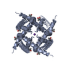
| Title | Structure of a K+ selective NaK mutant (NaK2K, Laue diffraction, no electric field) | ||||||||||||||||||||||||||||||
 Components Components | Potassium channel protein | ||||||||||||||||||||||||||||||
 Keywords Keywords | MEMBRANE PROTEIN / Potassium ion channel / EFX / electric field | ||||||||||||||||||||||||||||||
| Function / homology | Two pore domain potassium channel / Potassium channel domain / Ion channel / potassium channel activity / membrane / : / Potassium channel protein Function and homology information Function and homology information | ||||||||||||||||||||||||||||||
| Biological species |  Bacillus cereus m1550 (bacteria) Bacillus cereus m1550 (bacteria) | ||||||||||||||||||||||||||||||
| Method | X-RAY DIFFRACTION / SYNCHROTRON / MOLECULAR REPLACEMENT / Resolution: 2.01 Å | ||||||||||||||||||||||||||||||
 Authors Authors | Lee, B. / White, K.I. / Socolich, M.A. / Klureza, M.A. / Henning, R. / Srajer, V. / Ranganathan, R. / Hekstra, D. | ||||||||||||||||||||||||||||||
| Funding support |  United States, 9items United States, 9items
| ||||||||||||||||||||||||||||||
 Citation Citation | Journal: To Be Published Title: Direct visualization of electric field-stimulated ion conduction in a potassium channel Authors: Lee, B. / White, K.I. / Socolich, M.A. / Klureza, M.A. / Henning, R. / Srajer, V. / Ranganathan, R. / Hekstra, D. | ||||||||||||||||||||||||||||||
| History |
|
- Structure visualization
| Structure viewer | Molecule: MolmilJmol/JSmol |
|---|
- Downloads & links
-Download
| PDBx/mmCIF format | 8ctn.cif.gz | 112 KB | Display | PDBx/mmCIF format |
|---|---|---|---|---|
| PDB format | pdb8ctn.ent.gz | 89.5 KB | Display | PDB format |
| PDBx/mmJSON format | 8ctn.json.gz | Tree view | PDBx/mmJSON format | |
| Others |  Other downloads Other downloads |
-Validation report
| Arichive directory | https://data.pdbj.org/pub/pdb/validation_reports/ct/8ctnftp://data.pdbj.org/pub/pdb/validation_reports/ct/8ctn | HTTPS FTP |
|---|
-Related structure data
| Related structure data |  8ctsC  8cttC  8ctuC  8ctvC  8ctwC  8ctxC  8cu1C  8cu2C 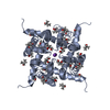 8cu3C 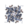 8cu4C C: citing same article ( |
|---|---|
| Similar structure data |
-Links
 PDBj
PDBj

- Assembly
| Deposited unit | 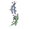
| |||||||||||||||||||||||||||||||||||||||
|---|---|---|---|---|---|---|---|---|---|---|---|---|---|---|---|---|---|---|---|---|---|---|---|---|---|---|---|---|---|---|---|---|---|---|---|---|---|---|---|---|
| 1 | 
| |||||||||||||||||||||||||||||||||||||||
| 2 | 
| |||||||||||||||||||||||||||||||||||||||
| Unit cell |
| |||||||||||||||||||||||||||||||||||||||
| Components on special symmetry positions |
|
-Components
| #1: Protein | Mass: 10640.477 Da / Num. of mol.: 2 Source method: isolated from a genetically manipulated source Source: (gene. exp.) Bacillus cereus m1550 (bacteria) / Gene: bcere0011_5790 / Production host: Escherichia coli (E. coli) / Strain (production host): SG13009 / References: UniProt: C2R3K4#2: Polysaccharide | alpha-D-glucopyranose-(1-4)-alpha-D-glucopyranose | / Mass: 342.297 Da / Num. of mol.: 1 / Source method: obtained synthetically#3: Chemical | ChemComp-MPD / ( 2-Methyl-2,4-pentanediol  Mass: 118.174 Da / Num. of mol.: 17 / Source method: obtained synthetically / Formula: C6H14O2 / Comment: precipitant*YM Mass: 118.174 Da / Num. of mol.: 17 / Source method: obtained synthetically / Formula: C6H14O2 / Comment: precipitant*YM#4: Chemical | ChemComp-K /   Mass: 39.098 Da / Num. of mol.: 12 / Source method: obtained synthetically / Formula: K / Feature type: SUBJECT OF INVESTIGATION Mass: 39.098 Da / Num. of mol.: 12 / Source method: obtained synthetically / Formula: K / Feature type: SUBJECT OF INVESTIGATION#5: Water | ChemComp-HOH / | Water Mass: 18.015 Da / Num. of mol.: 39 / Source method: isolated from a natural source / Formula: H2O Mass: 18.015 Da / Num. of mol.: 39 / Source method: isolated from a natural source / Formula: H2OHas ligand of interest | Y | |
|---|
-Experimental details
-Experiment
| Experiment | Method: X-RAY DIFFRACTION / Number of used crystals: 1 |
|---|
- Sample preparation
| Crystal | Density Matthews: 2.51 Å3/Da / Density % sol: 51.07 % |
|---|---|
| Crystal grow | Temperature: 293.15 K / Method: vapor diffusion, sitting drop Details: 100mM KCl, 200mM potassium citrate tribasic monohydrate, 100mM MES (pH 6.0 or 6.5), 56%-68% 2-methyl-2,4-pentanediol (MPD) |
-Data collection
| Diffraction | Mean temperature: 289 K / Ambient temp details: room temperature / Serial crystal experiment: N | |||||||||
|---|---|---|---|---|---|---|---|---|---|---|
| Diffraction source | Source: SYNCHROTRON / Site: APS / Beamline: 14-ID-B / Wavelength: 1.02-1.15 | |||||||||
| Detector | Type: RAYONIX MX340-HS / Detector: CCD / Date: Mar 21, 2020 | |||||||||
| Radiation | Protocol: LAUE / Monochromatic (M) / Laue (L): L / Scattering type: x-ray | |||||||||
| Radiation wavelength |
| |||||||||
| Reflection | Resolution: 2→100 Å / Num. obs: 10684 / % possible obs: 75.4 % / Redundancy: 3.7 % / Biso Wilson estimate: 18.3 Å2 / Rmerge(I) obs: 0.076 / Net I/σ(I): 42.5 | |||||||||
| Reflection shell | Resolution: 2→2.09 Å / Mean I/σ(I) obs: 7.67 / Num. unique obs: 562 / % possible all: 32.14 |
- Processing
| Software |
| |||||||||||||||||||||||||||||||||||
|---|---|---|---|---|---|---|---|---|---|---|---|---|---|---|---|---|---|---|---|---|---|---|---|---|---|---|---|---|---|---|---|---|---|---|---|---|
| Refinement | Method to determine structure: MOLECULAR REPLACEMENT Starting model: Room temperature NaK2K, monochromatic, SSRL Resolution: 2.01→21.42 Å / SU ML: 0.1765 / Cross valid method: FREE R-VALUE / σ(F): 2.99 / Phase error: 17.5854 Stereochemistry target values: GeoStd + Monomer Library + CDL v1.2
| |||||||||||||||||||||||||||||||||||
| Solvent computation | Shrinkage radii: 0.9 Å / VDW probe radii: 1.11 Å / Solvent model: FLAT BULK SOLVENT MODEL | |||||||||||||||||||||||||||||||||||
| Displacement parameters | Biso mean: 31.4 Å2 | |||||||||||||||||||||||||||||||||||
| Refinement step | Cycle: LAST / Resolution: 2.01→21.42 Å
| |||||||||||||||||||||||||||||||||||
| Refine LS restraints |
| |||||||||||||||||||||||||||||||||||
| LS refinement shell |
|
