 Movie
Movie Controller

[English] 日本語
 Yorodumi
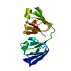
- PDB-7p53: Crystal Structure of Human gamma-D-crystallin mutant C110M at 1.5... -
+ Open data

- Basic information
| Entry | Database: PDB / ID: 7p53 | ||||||
|---|---|---|---|---|---|---|---|
| Title | Crystal Structure of Human gamma-D-crystallin mutant C110M at 1.57 Angstroms resolution | ||||||
 Components Components | Gamma-crystallin D | ||||||
 Keywords Keywords |  STRUCTURAL PROTEIN / Cysteine mutation / Recombinant / Eye lens STRUCTURAL PROTEIN / Cysteine mutation / Recombinant / Eye lens | ||||||
| Function / homology |  Function and homology information Function and homology informationlens fiber cell differentiation / structural constituent of eye lens / lens development in camera-type eye / visual perception / cellular response to reactive oxygen species / nucleus / cytoplasmSimilarity search - Function | ||||||
| Biological species |  Homo sapiens (human) Homo sapiens (human) | ||||||
| Method | X-RAY DIFFRACTION / SYNCHROTRON / MOLECULAR REPLACEMENT / Resolution: 1.57 Å | ||||||
 Authors Authors | Strofaldi, A. / Khan, A.R. / McManus, J. | ||||||
| Funding support |  Ireland, 1items Ireland, 1items
| ||||||
 Citation Citation | Journal: J.Mol.Biol. / Year: 2021 Title: Surface Exposed Free Cysteine Suppresses Crystallization of Human gamma D-Crystallin. Authors: Strofaldi, A. / Khan, A.R. / McManus, J.J. | ||||||
| History |
|
- Structure visualization
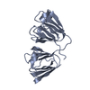
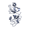
| Structure viewer | Molecule: MolmilJmol/JSmol |
|---|
- Downloads & links
-Download
| PDBx/mmCIF format | 7p53.cif.gz | 87.5 KB | Display | PDBx/mmCIF format |
|---|---|---|---|---|
| PDB format | pdb7p53.ent.gz | 61.1 KB | Display | PDB format |
| PDBx/mmJSON format | 7p53.json.gz | Tree view | PDBx/mmJSON format | |
| Others |  Other downloads Other downloads |
-Validation report
| Arichive directory | https://data.pdbj.org/pub/pdb/validation_reports/p5/7p53ftp://data.pdbj.org/pub/pdb/validation_reports/p5/7p53 | HTTPS FTP |
|---|
-Related structure data
| Related structure data | 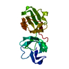 1hk0S S: Starting model for refinement |
|---|---|
| Similar structure data |


-Links
 PDBj
PDBj
- Assembly
| Deposited unit | 
| ||||||||||
|---|---|---|---|---|---|---|---|---|---|---|---|
| 1 |
| ||||||||||
| Unit cell |
|
-Components
| #1: Protein | Mass: 20663.021 Da / Num. of mol.: 1 / Mutation: C110M Source method: isolated from a genetically manipulated source Details: The protein is a mutant of Human gamma-D crystallin where the 110th cysteine is replaced by a methionine (C110M). Source: (gene. exp.) Homo sapiens (human) / Gene: CRYGD, CRYG4 / Organ: Eye / Production host:  Escherichia coli BL21(DE3) (bacteria) / Variant (production host): BL21-Gold(DE3) / References: UniProt: P07320 Escherichia coli BL21(DE3) (bacteria) / Variant (production host): BL21-Gold(DE3) / References: UniProt: P07320 |
|---|---|
| #2: Water | ChemComp-HOH / Water Mass: 18.015 Da / Num. of mol.: 108 / Source method: isolated from a natural source / Formula: H2O Mass: 18.015 Da / Num. of mol.: 108 / Source method: isolated from a natural source / Formula: H2O |
-Experimental details
-Experiment
| Experiment | Method: X-RAY DIFFRACTION / Number of used crystals: 1 |
|---|
- Sample preparation
| Crystal | Density Matthews: 2.05 Å3/Da / Density % sol: 40 % / Description: Rhombic crystals. |
|---|---|
| Crystal grow | Temperature: 277 K / Method: batch mode / pH: 7 Details: Crystals were obtained in 0.1M Sodium Phosphate Buffer pH 7 inducing LLPS at approximately 263K for 3hours from 150mg/mL solutions. Crystals were observed after few hours of incubation at 277K. |
-Data collection
| Diffraction | Mean temperature: 100 K / Serial crystal experiment: N |
|---|---|
| Diffraction source | Source: SYNCHROTRON / Site: SOLEIL  / Beamline: PROXIMA 2 / Wavelength: 0.97918 Å / Beamline: PROXIMA 2 / Wavelength: 0.97918 Å |
| Detector | Type: DECTRIS EIGER X 9M / Detector: PIXEL / Date: Mar 31, 2019 |
| Radiation | Protocol: SINGLE WAVELENGTH / Monochromatic (M) / Laue (L): M / Scattering type: x-ray |
| Radiation wavelength | Wavelength: 0.97918 Å / Relative weight: 1 |
| Reflection | Resolution: 1.57→52.84 Å / Num. obs: 22817 / % possible obs: 99.2 % / Redundancy: 6.1 % / Biso Wilson estimate: 25.8 Å2 / Rmerge(I) obs: 0.188 / Rpim(I) all: 0.083 / Rrim(I) all: 0.206 / Net I/σ(I): 6 |
| Reflection shell | Resolution: 1.57→1.6 Å / Redundancy: 5.3 % / Rmerge(I) obs: 1.941 / Mean I/σ(I) obs: 0.5 / Num. unique obs: 953 / Rpim(I) all: 0.883 / Rrim(I) all: 2.141 / % possible all: 85.8 |
- Processing
| Software |
| |||||||||||||||||||||||||||||||||||||||||||||||||||||||||||||||
|---|---|---|---|---|---|---|---|---|---|---|---|---|---|---|---|---|---|---|---|---|---|---|---|---|---|---|---|---|---|---|---|---|---|---|---|---|---|---|---|---|---|---|---|---|---|---|---|---|---|---|---|---|---|---|---|---|---|---|---|---|---|---|---|---|
| Refinement | Method to determine structure: MOLECULAR REPLACEMENT Starting model: 1hk0 Resolution: 1.57→45.51 Å / SU ML: 0.3044 / Cross valid method: THROUGHOUT / σ(F): 1.34 / Phase error: 35.562 Stereochemistry target values: GeoStd + Monomer Library + CDL v1.2
| |||||||||||||||||||||||||||||||||||||||||||||||||||||||||||||||
| Solvent computation | Shrinkage radii: 0.9 Å / VDW probe radii: 1.11 Å / Solvent model: FLAT BULK SOLVENT MODEL | |||||||||||||||||||||||||||||||||||||||||||||||||||||||||||||||
| Displacement parameters | Biso mean: 32.55 Å2 | |||||||||||||||||||||||||||||||||||||||||||||||||||||||||||||||
| Refinement step | Cycle: LAST / Resolution: 1.57→45.51 Å
| |||||||||||||||||||||||||||||||||||||||||||||||||||||||||||||||
| Refine LS restraints |
| |||||||||||||||||||||||||||||||||||||||||||||||||||||||||||||||
| LS refinement shell |
|
