 Movie
Movie Controller

[English] 日本語
 Yorodumi
- PDB-6eta: Crystal Structure of Human Gamma-D crystallin Mutant P23T+R36S at... -
+ Open data

- Basic information
| Entry | Database: PDB / ID: 6eta | ||||||
|---|---|---|---|---|---|---|---|
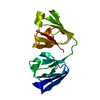
| Title | Crystal Structure of Human Gamma-D crystallin Mutant P23T+R36S at Room Temperature | ||||||
 Components Components | Gamma-crystallin D | ||||||
 Keywords Keywords |  STRUCTURAL PROTEIN / age-related cateract eye lens protein structural protein STRUCTURAL PROTEIN / age-related cateract eye lens protein structural protein | ||||||
| Function / homology |  Function and homology information Function and homology informationlens fiber cell differentiation / structural constituent of eye lens / lens development in camera-type eye / visual perception / cellular response to reactive oxygen species / nucleus / cytoplasmSimilarity search - Function | ||||||
| Biological species |  Homo sapiens (human) Homo sapiens (human) | ||||||
| Method | X-RAY DIFFRACTION / SYNCHROTRON / MOLECULAR REPLACEMENT / Resolution: 2.198 Å | ||||||
 Authors Authors | James, S. / McManus, J. / Khan, A.R. | ||||||
| Funding support |  Ireland, 1items Ireland, 1items
| ||||||
 Citation Citation | Journal: Biophys.J. / Year: 2019 Title: Temperature-Dependent Interactions Explain Normal and Inverted Solubility in a gamma D-Crystallin Mutant. Authors: Khan, A.R. / James, S. / Quinn, M.K. / Altan, I. / Charbonneau, P. / McManus, J.J. | ||||||
| History |
|
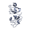
- Structure visualization
| Structure viewer | Molecule: MolmilJmol/JSmol |
|---|
- Downloads & links
-Download
| PDBx/mmCIF format | 6eta.cif.gz | 157.2 KB | Display | PDBx/mmCIF format |
|---|---|---|---|---|
| PDB format | pdb6eta.ent.gz | 124.1 KB | Display | PDB format |
| PDBx/mmJSON format | 6eta.json.gz | Tree view | PDBx/mmJSON format | |
| Others |  Other downloads Other downloads |
-Validation report
| Arichive directory | https://data.pdbj.org/pub/pdb/validation_reports/et/6etaftp://data.pdbj.org/pub/pdb/validation_reports/et/6eta | HTTPS FTP |
|---|
-Related structure data
| Related structure data | 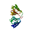 6etcC 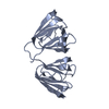 4jgfS S: Starting model for refinement C: citing same article ( |
|---|---|
| Similar structure data |


-Links
 PDBj
PDBj
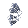
- Assembly
| Deposited unit | 
| ||||||||
|---|---|---|---|---|---|---|---|---|---|
| 1 | 
| ||||||||
| 2 | 
| ||||||||
| Unit cell |
|
-Components
| #1: Protein | Mass: 20700.039 Da / Num. of mol.: 2 Source method: isolated from a genetically manipulated source Source: (gene. exp.) Homo sapiens (human) / Gene: CRYGD, CRYG4 / Production host:  Escherichia coli (E. coli) / References: UniProt: P07320 Escherichia coli (E. coli) / References: UniProt: P07320#2: Water | ChemComp-HOH / | Water Mass: 18.015 Da / Num. of mol.: 112 / Source method: isolated from a natural source / Formula: H2O Mass: 18.015 Da / Num. of mol.: 112 / Source method: isolated from a natural source / Formula: H2O |
|---|
-Experimental details
-Experiment
| Experiment | Method: X-RAY DIFFRACTION / Number of used crystals: 1 |
|---|
- Sample preparation
| Crystal | Density Matthews: 2.85 Å3/Da / Density % sol: 56.79 % |
|---|---|
| Crystal grow | Temperature: 293 K / Method: batch mode / pH: 7 / Details: 0.1M phosphate, 20mM DTT |
-Data collection
| Diffraction | Mean temperature: 100 K |
|---|---|
| Diffraction source | Source: SYNCHROTRON / Site: SOLEIL  / Beamline: PROXIMA 2 / Wavelength: 0.9786 Å / Beamline: PROXIMA 2 / Wavelength: 0.9786 Å |
| Detector | Type: ADSC QUANTUM 315 / Detector: CCD / Date: Jul 16, 2015 |
| Radiation | Protocol: SINGLE WAVELENGTH / Monochromatic (M) / Laue (L): M / Scattering type: x-ray |
| Radiation wavelength | Wavelength: 0.9786 Å / Relative weight: 1 |
| Reflection | Resolution: 2.198→48.165 Å / Num. obs: 24656 / % possible obs: 99.63 % / Redundancy: 6.7 % / Rmerge(I) obs: 0.129 / Rrim(I) all: 0.139 / Net I/σ(I): 9.2 |
| Reflection shell | Resolution: 2.198→2.27 Å / Rmerge(I) obs: 1.29 / Rrim(I) all: 1.43 |
- Processing
| Software |
| ||||||||||||||||||||||||||||||||||||||||||||||||||||||||||||||||||||||
|---|---|---|---|---|---|---|---|---|---|---|---|---|---|---|---|---|---|---|---|---|---|---|---|---|---|---|---|---|---|---|---|---|---|---|---|---|---|---|---|---|---|---|---|---|---|---|---|---|---|---|---|---|---|---|---|---|---|---|---|---|---|---|---|---|---|---|---|---|---|---|---|
| Refinement | Method to determine structure: MOLECULAR REPLACEMENT Starting model: 4jgf Resolution: 2.198→48.165 Å / SU ML: 0.31 / Cross valid method: FREE R-VALUE / σ(F): 1.34 / Phase error: 28.36
| ||||||||||||||||||||||||||||||||||||||||||||||||||||||||||||||||||||||
| Solvent computation | Shrinkage radii: 0.9 Å / VDW probe radii: 1.11 Å | ||||||||||||||||||||||||||||||||||||||||||||||||||||||||||||||||||||||
| Refinement step | Cycle: LAST / Resolution: 2.198→48.165 Å
| ||||||||||||||||||||||||||||||||||||||||||||||||||||||||||||||||||||||
| Refine LS restraints |
| ||||||||||||||||||||||||||||||||||||||||||||||||||||||||||||||||||||||
| LS refinement shell |
| ||||||||||||||||||||||||||||||||||||||||||||||||||||||||||||||||||||||
| Refinement TLS params. | Method: refined / Origin x: 5.6744 Å / Origin y: 91.3059 Å / Origin z: -7.3998 Å
| ||||||||||||||||||||||||||||||||||||||||||||||||||||||||||||||||||||||
| Refinement TLS group | Selection details: all |
