 Movie
Movie Controller


+ Open data

- Basic information
| Entry | Database: PDB / ID: 6l6m | ||||||
|---|---|---|---|---|---|---|---|
| Title | HSP18.5 from E. histolytica | ||||||
 Components Components | Heat shock protein hsp20 family putative Heat shock response Heat shock response | ||||||
 Keywords Keywords | CHAPERONE / Small heat shock protein HSP18.5 Molecular chaperone E. histolytica | ||||||
| Function / homology | Hsp20/alpha crystallin family / Small heat shock protein (sHSP) domain profile. / Alpha crystallin/Hsp20 domain / HSP20-like chaperone / Heat shock protein, Hsp20 family, putative Function and homology information Function and homology information | ||||||
| Biological species |   Entamoeba histolytica (eukaryote) Entamoeba histolytica (eukaryote) | ||||||
| Method | X-RAY DIFFRACTION / SYNCHROTRON / MOLECULAR REPLACEMENT / Resolution: 3.28000381979 Å | ||||||
 Authors Authors | Kurre, D. / Suguna, K. | ||||||
| Funding support |  India, 1items India, 1items
| ||||||
 Citation Citation | Journal: Proteins / Year: 2021 Title: Network of Entamoeba histolytica HSP18.5 dimers formed by two overlapping [IV]-X-[IV] motifs. Authors: Kurre, D. / Suguna, K. | ||||||
| History |
|
- Structure visualization
| Structure viewer | Molecule: MolmilJmol/JSmol |
|---|
- Downloads & links
-Download
| PDBx/mmCIF format | 6l6m.cif.gz | 116.3 KB | Display | PDBx/mmCIF format |
|---|---|---|---|---|
| PDB format | pdb6l6m.ent.gz | 71.6 KB | Display | PDB format |
| PDBx/mmJSON format | 6l6m.json.gz | Tree view | PDBx/mmJSON format | |
| Others |  Other downloads Other downloads |
-Validation report
| Arichive directory | https://data.pdbj.org/pub/pdb/validation_reports/l6/6l6mftp://data.pdbj.org/pub/pdb/validation_reports/l6/6l6m | HTTPS FTP |
|---|
-Related structure data
| Related structure data | 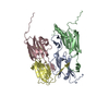 3w1zS S: Starting model for refinement |
|---|---|
| Similar structure data |


-Links
 PDBj
PDBj


- Assembly
| Deposited unit | 
| ||||||||||||
|---|---|---|---|---|---|---|---|---|---|---|---|---|---|
| 1 | 
| ||||||||||||
| Unit cell |
|
-Components
| #1: Protein | Heat shock response / Heat shock protein / Hsp20 family / putative Mass: 18848.393 Da / Num. of mol.: 4 Source method: isolated from a genetically manipulated source Source: (gene. exp.) Entamoeba histolytica (eukaryote) / Gene: CL6EHI_193390, EHI_193390 / Production host:  Escherichia coli (E. coli) / References: UniProt: C4M4U3 Escherichia coli (E. coli) / References: UniProt: C4M4U3 |
|---|
-Experimental details
-Experiment
| Experiment | Method: X-RAY DIFFRACTION / Number of used crystals: 1 |
|---|
- Sample preparation
| Crystal | Density Matthews: 3.32 Å3/Da / Density % sol: 65.92 % |
|---|---|
| Crystal grow | Temperature: 295 K / Method: vapor diffusion, hanging drop / pH: 6.2 Details: 0.1 M Bis-Tris 160 mM ammonium acetate 45% 2-Methyl-2,4-pentanediol PH range: 6.0-7.5 |
-Data collection
| Diffraction | Mean temperature: 100 K / Serial crystal experiment: N |
|---|---|
| Diffraction source | Source: SYNCHROTRON / Site: ESRF  / Beamline: ID30B / Wavelength: 0.991872 Å / Beamline: ID30B / Wavelength: 0.991872 Å |
| Detector | Type: DECTRIS PILATUS 6M / Detector: PIXEL / Date: Aug 31, 2017 |
| Radiation | Protocol: SINGLE WAVELENGTH / Monochromatic (M) / Laue (L): M / Scattering type: x-ray |
| Radiation wavelength | Wavelength: 0.991872 Å / Relative weight: 1 |
| Reflection | Resolution: 3.28→79.53 Å / Num. obs: 15560 / % possible obs: 99.9 % / Redundancy: 11.1 % / Biso Wilson estimate: 112.375814491 Å2 / CC1/2: 0.99 / Net I/σ(I): 5.99 |
| Reflection shell | Resolution: 3.28→3.43 Å / Num. unique obs: 2004 / CC1/2: 0.99 / % possible all: 99.9 |
- Processing
| Software |
| ||||||||||||||||||||||||||||||||||||||||||
|---|---|---|---|---|---|---|---|---|---|---|---|---|---|---|---|---|---|---|---|---|---|---|---|---|---|---|---|---|---|---|---|---|---|---|---|---|---|---|---|---|---|---|---|
| Refinement | Method to determine structure: MOLECULAR REPLACEMENT Starting model: 3W1Z Resolution: 3.28000381979→79.529 Å / SU ML: 0.359075090824 / Cross valid method: FREE R-VALUE / σ(F): 1.91706635515 / Phase error: 24.653739223 Stereochemistry target values: GeoStd + Monomer Library + CDL v1.2
| ||||||||||||||||||||||||||||||||||||||||||
| Solvent computation | Shrinkage radii: 0.9 Å / VDW probe radii: 1.11 Å / Solvent model: FLAT BULK SOLVENT MODEL | ||||||||||||||||||||||||||||||||||||||||||
| Displacement parameters | Biso mean: 99.2352389117 Å2 | ||||||||||||||||||||||||||||||||||||||||||
| Refinement step | Cycle: LAST / Resolution: 3.28000381979→79.529 Å
| ||||||||||||||||||||||||||||||||||||||||||
| Refine LS restraints |
| ||||||||||||||||||||||||||||||||||||||||||
| LS refinement shell |
|
