 Movie
Movie Controller


+ Open data

- Basic information
| Entry | Database: PDB / ID: 3w1z | ||||||
|---|---|---|---|---|---|---|---|
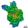
| Title | Heat shock protein 16.0 from Schizosaccharomyces pombe | ||||||
 Components Components | Heat shock protein 16 Heat shock response Heat shock response | ||||||
 Keywords Keywords | CHAPERONE / alpha-crystallin domain / small heat shock protein | ||||||
| Function / homology |  Function and homology information Function and homology informationprotein aggregate center / cellular response to misfolded protein / cellular response to unfolded protein / chaperone-mediated protein folding / unfolded protein binding / cellular response to heat / identical protein binding / nucleus / cytosol / cytoplasmSimilarity search - Function | ||||||
| Biological species |  Schizosaccharomyces pombe (fission yeast) Schizosaccharomyces pombe (fission yeast) | ||||||
| Method | X-RAY DIFFRACTION / SYNCHROTRON / MOLECULAR REPLACEMENT / Resolution: 2.401 Å | ||||||
 Authors Authors | Hanazono, Y. / Takeda, K. / Akiyama, N. / Aikawa, Y. / Miki, K. | ||||||
 Citation Citation | Journal: Structure / Year: 2013 Title: Nonequivalence Observed for the 16-Meric Structure of a Small Heat Shock Protein, SpHsp16.0, from Schizosaccharomyces pombe Authors: Hanazono, Y. / Takeda, K. / Oka, T. / Abe, T. / Tomonari, T. / Akiyama, N. / Aikawa, Y. / Yohda, M. / Miki, K. | ||||||
| History |
|
- Structure visualization
| Structure viewer | Molecule: MolmilJmol/JSmol |
|---|
- Downloads & links
-Download
| PDBx/mmCIF format | 3w1z.cif.gz | 113.1 KB | Display | PDBx/mmCIF format |
|---|---|---|---|---|
| PDB format | pdb3w1z.ent.gz | 90.2 KB | Display | PDB format |
| PDBx/mmJSON format | 3w1z.json.gz | Tree view | PDBx/mmJSON format | |
| Others |  Other downloads Other downloads |
-Validation report
| Arichive directory | https://data.pdbj.org/pub/pdb/validation_reports/w1/3w1zftp://data.pdbj.org/pub/pdb/validation_reports/w1/3w1z | HTTPS FTP |
|---|
-Related structure data
| Related structure data |  1gmeS S: Starting model for refinement |
|---|---|
| Similar structure data |


-Links
 PDBj
PDBj


- Assembly
| Deposited unit | 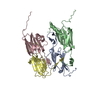
| ||||||||
|---|---|---|---|---|---|---|---|---|---|
| 1 | 
| ||||||||
| Unit cell |
|
-Components
| #1: Protein | Heat shock response / 16 kDa heat shock protein Mass: 15987.883 Da / Num. of mol.: 4 Source method: isolated from a genetically manipulated source Source: (gene. exp.) Schizosaccharomyces pombe (fission yeast)Strain: 972 / Gene: hsp16 / Production host:  Escherichia coli (E. coli) / References: UniProt: O14368 Escherichia coli (E. coli) / References: UniProt: O14368#2: Water | ChemComp-HOH / | Water Mass: 18.015 Da / Num. of mol.: 93 / Source method: isolated from a natural source / Formula: H2O Mass: 18.015 Da / Num. of mol.: 93 / Source method: isolated from a natural source / Formula: H2O |
|---|
-Experimental details
-Experiment
| Experiment | Method: X-RAY DIFFRACTION / Number of used crystals: 1 |
|---|
- Sample preparation
| Crystal | Density Matthews: 2.27 Å3/Da / Density % sol: 45.85 % |
|---|---|
| Crystal grow | Temperature: 293 K / Method: vapor diffusion, sitting drop / pH: 9 Details: 15% PEGMME550, 100mM sodium chloride, 100mM Bicine buffer, pH 9.0, VAPOR DIFFUSION, SITTING DROP, temperature 293K |
-Data collection
| Diffraction | Mean temperature: 100 K |
|---|---|
| Diffraction source | Source: SYNCHROTRON / Site: SPring-8  / Beamline: BL41XU / Wavelength: 1 Å / Beamline: BL41XU / Wavelength: 1 Å |
| Detector | Type: ADSC QUANTUM 315 / Detector: CCD / Date: Oct 27, 2005 |
| Radiation | Protocol: SINGLE WAVELENGTH / Monochromatic (M) / Laue (L): M / Scattering type: x-ray |
| Radiation wavelength | Wavelength: 1 Å / Relative weight: 1 |
| Reflection | Resolution: 2.4→50 Å / Num. obs: 22653 / % possible obs: 97.8 % / Redundancy: 5.3 % / Biso Wilson estimate: 40.13 Å2 / Rsym value: 0.043 / Net I/σ(I): 21.4 |
| Reflection shell | Resolution: 2.4→2.49 Å / Redundancy: 3.7 % / Mean I/σ(I) obs: 2.1 / Rsym value: 0.254 / % possible all: 95.6 |
- Processing
| Software |
| |||||||||||||||||||||||||||||||||||||||||||||||||||||||||||||||
|---|---|---|---|---|---|---|---|---|---|---|---|---|---|---|---|---|---|---|---|---|---|---|---|---|---|---|---|---|---|---|---|---|---|---|---|---|---|---|---|---|---|---|---|---|---|---|---|---|---|---|---|---|---|---|---|---|---|---|---|---|---|---|---|---|
| Refinement | Method to determine structure: MOLECULAR REPLACEMENT Starting model: 1GME Resolution: 2.401→44.834 Å / Occupancy max: 1 / Occupancy min: 0.45 / FOM work R set: 0.7417 / SU ML: 0.41 / σ(F): 0 / Phase error: 31.84 / Stereochemistry target values: ML
| |||||||||||||||||||||||||||||||||||||||||||||||||||||||||||||||
| Solvent computation | Shrinkage radii: 0.9 Å / VDW probe radii: 1.11 Å / Solvent model: FLAT BULK SOLVENT MODEL | |||||||||||||||||||||||||||||||||||||||||||||||||||||||||||||||
| Displacement parameters | Biso max: 137.45 Å2 / Biso mean: 51.4625 Å2 / Biso min: 10.29 Å2 | |||||||||||||||||||||||||||||||||||||||||||||||||||||||||||||||
| Refinement step | Cycle: LAST / Resolution: 2.401→44.834 Å
| |||||||||||||||||||||||||||||||||||||||||||||||||||||||||||||||
| Refine LS restraints |
| |||||||||||||||||||||||||||||||||||||||||||||||||||||||||||||||
| LS refinement shell | Refine-ID: X-RAY DIFFRACTION / Total num. of bins used: 8
|
