 Movie
Movie Controller

[English] 日本語
 Yorodumi
- PDB-5r4p: PanDDA analysis group deposition -- Crystal Structure of HUMAN CL... -
+ Open data

- Basic information
| Entry | Database: PDB / ID: 5r4p | ||||||
|---|---|---|---|---|---|---|---|
| Title | PanDDA analysis group deposition -- Crystal Structure of HUMAN CLEAVAGE FACTOR IM in complex with NM450-1 | ||||||
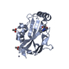
 Components Components | Cleavage and polyadenylation specificity factor subunit 5 | ||||||
 Keywords Keywords | RNA BINDING PROTEIN / PanDDA / SGC - Diamond I04-1 fragment screening / NUDIX domain / XChemExplorer / CPSF5 / RNA PROCESSING / CLEAVAGE FACTOR / MRNA PROCESSING / NUCLEUS / PHOSPHOPROTEIN / RNA-BINDING | ||||||
| Function / homology |  Function and homology information Function and homology informationpositive regulation of pro-B cell differentiation / : / mRNA cleavage factor complex / Processing of Intronless Pre-mRNAs / positive regulation of stem cell differentiation / mRNA cleavage and polyadenylation specificity factor complex / mRNA alternative polyadenylation / mRNA 3'-UTR AU-rich region binding / paraspeckles / mRNA 3'-end processing ...positive regulation of pro-B cell differentiation / : / mRNA cleavage factor complex / Processing of Intronless Pre-mRNAs / positive regulation of stem cell differentiation / mRNA cleavage and polyadenylation specificity factor complex / mRNA alternative polyadenylation / mRNA 3'-UTR AU-rich region binding / paraspeckles / mRNA 3'-end processing / mRNA 3'-end processing / protein heterotetramerization / RNA Polymerase II Transcription Termination / post-transcriptional regulation of gene expression / : / Processing of Capped Intron-Containing Pre-mRNA / centriolar satellite / protein tetramerization / mRNA processing / histone deacetylase binding / cell differentiation / nuclear body / mRNA binding / centrosome / chromatin binding / protein homodimerization activity / RNA binding / nucleoplasm / identical protein binding / nucleus / cytoplasmSimilarity search - Function | ||||||
| Biological species |  Homo sapiens (human) Homo sapiens (human) | ||||||
| Method | X-RAY DIFFRACTION / SYNCHROTRON / FOURIER SYNTHESIS / molecular replacement / Resolution: 1.78 Å | ||||||
 Authors Authors | Kidd, S.L. / Mateu, N. / Talon, R. / Krojer, T. / Aimon, A. / Bradley, A.R. / Fairhead, M. / Diaz-Saez, L. / Sore, H.F. / Madin, A. ...Kidd, S.L. / Mateu, N. / Talon, R. / Krojer, T. / Aimon, A. / Bradley, A.R. / Fairhead, M. / Diaz-Saez, L. / Sore, H.F. / Madin, A. / Huber, K.V.M. / von Delft, F. / Spring, D.R. | ||||||
 Citation Citation | Journal: To Be Published Title: PanDDA analysis group deposition Authors: Kidd, S.L. / Mateu, N. / Talon, R. / Krojer, T. / Aimon, A. / Bradley, A.R. / Fairhead, M. / Diaz-Saez, L. / Sore, H.F. / Madin, A. / Huber, K.V.M. / von Delft, F. / Spring, D.R. | ||||||
| History |
|
- Structure visualization
| Structure viewer | Molecule: MolmilJmol/JSmol |
|---|
- Downloads & links
-Download
| PDBx/mmCIF format | 5r4p.cif.gz | 100.7 KB | Display | PDBx/mmCIF format |
|---|---|---|---|---|
| PDB format | pdb5r4p.ent.gz | 76 KB | Display | PDB format |
| PDBx/mmJSON format | 5r4p.json.gz | Tree view | PDBx/mmJSON format | |
| Others |  Other downloads Other downloads |
-Validation report
| Arichive directory | https://data.pdbj.org/pub/pdb/validation_reports/r4/5r4pftp://data.pdbj.org/pub/pdb/validation_reports/r4/5r4p | HTTPS FTP |
|---|
-Group deposition
| ID | G_1002126 (6 entries) |
|---|---|
| Title | PanDDA analysis group deposition |
| Type | changed state |
| Description | HUMAN CLEAVAGE FACTOR IM screened against the DOS fragment library by X-ray Crystallography at the XChem facility of Diamond Light Source beamline I04-1 |


-Related structure data
| Related structure data |  3bapS S: Starting model for refinement |
|---|---|
| Similar structure data |


-Links
 PDBj
PDBj

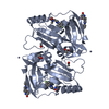
- Assembly
| Deposited unit | 
| ||||||||
|---|---|---|---|---|---|---|---|---|---|
| 1 | 
| ||||||||
| 2 | 
| ||||||||
| Unit cell |
|
-Components
| #1: Protein | / Cleavage and polyadenylation specificity factor 25 kDa subunit / CPSF 25 kDa subunit / Cleavage ...Cleavage and polyadenylation specificity factor 25 kDa subunit / CPSF 25 kDa subunit / Cleavage factor Im complex 25 kDa subunit / CFIm25 / Nucleoside diphosphate-linked moiety X motif 21 / Nudix motif 21 / Nudix hydrolase 21 / Pre-mRNA cleavage factor Im 68 kDa subunit Mass: 22874.393 Da / Num. of mol.: 2 Source method: isolated from a genetically manipulated source Source: (gene. exp.) Homo sapiens (human) / Gene: NUDT21, CFIM25, CPSF25, CPSF5 / Production host:  Escherichia coli (E. coli) / References: UniProt: O43809 Escherichia coli (E. coli) / References: UniProt: O43809#2: Chemical | ChemComp-ZN /   Mass: 65.409 Da / Num. of mol.: 6 / Source method: obtained synthetically / Formula: Zn Mass: 65.409 Da / Num. of mol.: 6 / Source method: obtained synthetically / Formula: Zn#3: Chemical | ChemComp-ACT / | Acetate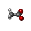  Mass: 59.044 Da / Num. of mol.: 1 / Source method: obtained synthetically / Formula: C2H3O2 Mass: 59.044 Da / Num. of mol.: 1 / Source method: obtained synthetically / Formula: C2H3O2#4: Chemical | ChemComp-RVY / ( |   Mass: 270.287 Da / Num. of mol.: 1 / Source method: obtained synthetically / Formula: C14H14N4O2 / Feature type: SUBJECT OF INVESTIGATION Mass: 270.287 Da / Num. of mol.: 1 / Source method: obtained synthetically / Formula: C14H14N4O2 / Feature type: SUBJECT OF INVESTIGATION#5: Water | ChemComp-HOH / | Water Mass: 18.015 Da / Num. of mol.: 255 / Source method: isolated from a natural source / Formula: H2O Mass: 18.015 Da / Num. of mol.: 255 / Source method: isolated from a natural source / Formula: H2OHas ligand of interest | Y | |
|---|
-Experimental details
-Experiment
| Experiment | Method: X-RAY DIFFRACTION / Number of used crystals: 1 |
|---|
- Sample preparation
| Crystal | Density Matthews: 2.24 Å3/Da / Density % sol: 45.07 % / Mosaicity: 0 ° |
|---|---|
| Crystal grow | Temperature: 277 K / Method: vapor diffusion, sitting drop / pH: 5.1 / Details: 0.1M acetate pH 5.1, 0.0025M ZnAC, 6% PEG3K |
-Data collection
| Diffraction | Mean temperature: 100 K | ||||||||||||||||||||||||||||||
|---|---|---|---|---|---|---|---|---|---|---|---|---|---|---|---|---|---|---|---|---|---|---|---|---|---|---|---|---|---|---|---|
| Diffraction source | Source: SYNCHROTRON / Site: Diamond  / Beamline: I04-1 / Wavelength: 0.92819 Å / Beamline: I04-1 / Wavelength: 0.92819 Å | ||||||||||||||||||||||||||||||
| Detector | Type: DECTRIS PILATUS 6M / Detector: PIXEL / Date: Jul 7, 2017 | ||||||||||||||||||||||||||||||
| Radiation | Protocol: SINGLE WAVELENGTH / Scattering type: x-ray | ||||||||||||||||||||||||||||||
| Radiation wavelength | Wavelength: 0.92819 Å / Relative weight: 1 | ||||||||||||||||||||||||||||||
| Reflection | Resolution: 1.78→51.68 Å / Num. obs: 43715 / % possible obs: 100 % / Redundancy: 9.6 % / CC1/2: 0.999 / Rmerge(I) obs: 0.128 / Rpim(I) all: 0.043 / Rrim(I) all: 0.135 / Net I/σ(I): 12.4 / Num. measured all: 421169 / Scaling rejects: 0 | ||||||||||||||||||||||||||||||
| Reflection shell | Diffraction-ID: 1
|
-Phasing
| Phasing | Method: molecular replacement |
|---|
- Processing
| Software |
| |||||||||||||||||||||||||||||||||||||||||||||||||||||||||||||||||||||||||||
|---|---|---|---|---|---|---|---|---|---|---|---|---|---|---|---|---|---|---|---|---|---|---|---|---|---|---|---|---|---|---|---|---|---|---|---|---|---|---|---|---|---|---|---|---|---|---|---|---|---|---|---|---|---|---|---|---|---|---|---|---|---|---|---|---|---|---|---|---|---|---|---|---|---|---|---|---|
| Refinement | Method to determine structure: FOURIER SYNTHESIS Starting model: 3BAP Resolution: 1.78→51.74 Å / Cor.coef. Fo:Fc: 0.957 / Cor.coef. Fo:Fc free: 0.942 / SU B: 4.613 / SU ML: 0.134 / Cross valid method: THROUGHOUT / σ(F): 0 / ESU R: 0.168 / ESU R Free: 0.148 / Stereochemistry target values: MAXIMUM LIKELIHOOD Details: HYDROGENS HAVE BEEN ADDED IN THE RIDING POSITIONS U VALUES : REFINED INDIVIDUALLY
| |||||||||||||||||||||||||||||||||||||||||||||||||||||||||||||||||||||||||||
| Solvent computation | Ion probe radii: 0.8 Å / Shrinkage radii: 0.8 Å / VDW probe radii: 1.2 Å / Solvent model: MASK | |||||||||||||||||||||||||||||||||||||||||||||||||||||||||||||||||||||||||||
| Displacement parameters | Biso max: 159.68 Å2 / Biso mean: 35.519 Å2 / Biso min: 17.4 Å2
| |||||||||||||||||||||||||||||||||||||||||||||||||||||||||||||||||||||||||||
| Refinement step | Cycle: final / Resolution: 1.78→51.74 Å
| |||||||||||||||||||||||||||||||||||||||||||||||||||||||||||||||||||||||||||
| Refine LS restraints |
| |||||||||||||||||||||||||||||||||||||||||||||||||||||||||||||||||||||||||||
| LS refinement shell | Resolution: 1.78→1.826 Å / Total num. of bins used: 20
|
