 Movie
Movie Controller

[English] 日本語
 Yorodumi
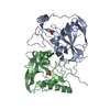
- PDB-3bap: Crystal Structure of the 25 kDa Subunit of Human Cleavage Factor Im -
+ Open data

- Basic information
| Entry | Database: PDB / ID: 3bap | ||||||
|---|---|---|---|---|---|---|---|
| Title | Crystal Structure of the 25 kDa Subunit of Human Cleavage Factor Im | ||||||
 Components Components | Cleavage and polyadenylation specificity factor subunit 5 | ||||||
 Keywords Keywords | RNA BINDING PROTEIN / CPSF5 / RNA processing / cleavage factor / mRNA processing / Nucleus / Phosphoprotein / RNA-binding | ||||||
| Function / homology |  Function and homology information Function and homology informationpositive regulation of pro-B cell differentiation / : / mRNA cleavage factor complex / Processing of Intronless Pre-mRNAs / positive regulation of stem cell differentiation / mRNA cleavage and polyadenylation specificity factor complex / mRNA alternative polyadenylation / mRNA 3'-UTR AU-rich region binding / paraspeckles / mRNA 3'-end processing ...positive regulation of pro-B cell differentiation / : / mRNA cleavage factor complex / Processing of Intronless Pre-mRNAs / positive regulation of stem cell differentiation / mRNA cleavage and polyadenylation specificity factor complex / mRNA alternative polyadenylation / mRNA 3'-UTR AU-rich region binding / paraspeckles / mRNA 3'-end processing / mRNA 3'-end processing / protein heterotetramerization / RNA Polymerase II Transcription Termination / post-transcriptional regulation of gene expression / : / Processing of Capped Intron-Containing Pre-mRNA / centriolar satellite / protein tetramerization / mRNA processing / histone deacetylase binding / cell differentiation / nuclear body / mRNA binding / centrosome / chromatin binding / protein homodimerization activity / RNA binding / nucleoplasm / identical protein binding / nucleus / cytoplasmSimilarity search - Function | ||||||
| Biological species |  Homo sapiens (human) Homo sapiens (human) | ||||||
| Method | X-RAY DIFFRACTION / SYNCHROTRON / Resolution: 1.85 Å | ||||||
 Authors Authors | Coseno, M. / Doublie, S. | ||||||
 Citation Citation | Journal: Nucleic Acids Res. / Year: 2008 Title: Crystal structure of the 25 kDa subunit of human cleavage factor Im. Authors: Coseno, M. / Martin, G. / Berger, C. / Gilmartin, G. / Keller, W. / Doublie, S. | ||||||
| History |
|
- Structure visualization
| Structure viewer | Molecule: MolmilJmol/JSmol |
|---|
- Downloads & links
-Download
| PDBx/mmCIF format | 3bap.cif.gz | 54.7 KB | Display | PDBx/mmCIF format |
|---|---|---|---|---|
| PDB format | pdb3bap.ent.gz | 41.8 KB | Display | PDB format |
| PDBx/mmJSON format | 3bap.json.gz | Tree view | PDBx/mmJSON format | |
| Others |  Other downloads Other downloads |
-Validation report
| Arichive directory | https://data.pdbj.org/pub/pdb/validation_reports/ba/3bapftp://data.pdbj.org/pub/pdb/validation_reports/ba/3bap | HTTPS FTP |
|---|
-Related structure data


-Links
 PDBj
PDBj

- Assembly
| Deposited unit | 
| ||||||||
|---|---|---|---|---|---|---|---|---|---|
| 1 | 
| ||||||||
| Unit cell |
|
-Components
| #1: Protein | / Cleavage and polyadenylation specificity factor 25 kDa subunit / CPSF 25 kDa subunit / Pre-mRNA ...Cleavage and polyadenylation specificity factor 25 kDa subunit / CPSF 25 kDa subunit / Pre-mRNA cleavage factor Im 25 kDa subunit / Nucleoside diphosphate-linked moiety X motif 21 / Nudix motif 21 Mass: 26446.824 Da / Num. of mol.: 1 Source method: isolated from a genetically manipulated source Source: (gene. exp.) Homo sapiens (human) / Gene: NUDT21, CFIM25, CPSF25, CPSF5 / Production host:  Escherichia coli (E. coli) / Strain (production host): Rosetta (DE3) pLysS / References: UniProt: O43809 Escherichia coli (E. coli) / Strain (production host): Rosetta (DE3) pLysS / References: UniProt: O43809 |
|---|---|
| #2: Chemical | ChemComp-GOL / Glycerol  Mass: 92.094 Da / Num. of mol.: 1 / Source method: obtained synthetically / Formula: C3H8O3 Mass: 92.094 Da / Num. of mol.: 1 / Source method: obtained synthetically / Formula: C3H8O3 |
| #3: Water | ChemComp-HOH / Water Mass: 18.015 Da / Num. of mol.: 168 / Source method: isolated from a natural source / Formula: H2O Mass: 18.015 Da / Num. of mol.: 168 / Source method: isolated from a natural source / Formula: H2O |
-Experimental details
-Experiment
| Experiment | Method: X-RAY DIFFRACTION / Number of used crystals: 1 |
|---|
- Sample preparation
| Crystal | Density Matthews: 2.53 Å3/Da / Density % sol: 51.37 % |
|---|---|
| Crystal grow | Temperature: 285 K / Method: vapor diffusion, hanging drop / pH: 8.5 Details: 25% PEG 3350, 0.2M Magnesium chloride, 0.1M Tris-HCl pH 8.5, VAPOR DIFFUSION, HANGING DROP, temperature 285K |
-Data collection
| Diffraction | Mean temperature: 100 K | |||||||||||||||||||||||||||||||||||||||||||||||||||||||||||||||||||||||||||||
|---|---|---|---|---|---|---|---|---|---|---|---|---|---|---|---|---|---|---|---|---|---|---|---|---|---|---|---|---|---|---|---|---|---|---|---|---|---|---|---|---|---|---|---|---|---|---|---|---|---|---|---|---|---|---|---|---|---|---|---|---|---|---|---|---|---|---|---|---|---|---|---|---|---|---|---|---|---|---|
| Diffraction source | Source: SYNCHROTRON / Site: APS  / Beamline: 23-ID-D / Wavelength: 0.97923, 0.97939, 0.97166 / Beamline: 23-ID-D / Wavelength: 0.97923, 0.97939, 0.97166 | |||||||||||||||||||||||||||||||||||||||||||||||||||||||||||||||||||||||||||||
| Detector | Type: MARMOSAIC 300 mm CCD / Detector: CCD / Date: Aug 3, 2006 / Details: mirrors | |||||||||||||||||||||||||||||||||||||||||||||||||||||||||||||||||||||||||||||
| Radiation | Monochromator: DCM - PAIR OF FLAT SI CRYSTALS / Protocol: MAD / Monochromatic (M) / Laue (L): M / Scattering type: x-ray | |||||||||||||||||||||||||||||||||||||||||||||||||||||||||||||||||||||||||||||
| Radiation wavelength |
| |||||||||||||||||||||||||||||||||||||||||||||||||||||||||||||||||||||||||||||
| Reflection | Redundancy: 6.6 % / Av σ(I) over netI: 14.2 / Number: 148933 / Rmerge(I) obs: 0.073 / Χ2: 1.05 / D res high: 1.85 Å / D res low: 30 Å / Num. obs: 22468 / % possible obs: 96.5 | |||||||||||||||||||||||||||||||||||||||||||||||||||||||||||||||||||||||||||||
| Diffraction reflection shell |
| |||||||||||||||||||||||||||||||||||||||||||||||||||||||||||||||||||||||||||||
| Reflection | Resolution: 1.82→30 Å / Num. all: 23255 / Num. obs: 22805 / % possible obs: 98.1 % / Redundancy: 6.8 % / Biso Wilson estimate: 31.752 Å2 / Rmerge(I) obs: 0.086 / Net I/σ(I): 16.09 | |||||||||||||||||||||||||||||||||||||||||||||||||||||||||||||||||||||||||||||
| Reflection shell | Resolution: 1.82→1.89 Å / Redundancy: 5.6 % / Rmerge(I) obs: 0.0517 / Mean I/σ(I) obs: 2.2 |
- Processing
| Software |
| ||||||||||||||||||||||||||||||||||||||||||||||||||||||||||||||||||||||||||||||||
|---|---|---|---|---|---|---|---|---|---|---|---|---|---|---|---|---|---|---|---|---|---|---|---|---|---|---|---|---|---|---|---|---|---|---|---|---|---|---|---|---|---|---|---|---|---|---|---|---|---|---|---|---|---|---|---|---|---|---|---|---|---|---|---|---|---|---|---|---|---|---|---|---|---|---|---|---|---|---|---|---|---|
| Refinement | Resolution: 1.85→30 Å / σ(F): 0 / Stereochemistry target values: Engh & Huber
| ||||||||||||||||||||||||||||||||||||||||||||||||||||||||||||||||||||||||||||||||
| Solvent computation | Bsol: 38.14 Å2 | ||||||||||||||||||||||||||||||||||||||||||||||||||||||||||||||||||||||||||||||||
| Displacement parameters | Biso mean: 38.783 Å2
| ||||||||||||||||||||||||||||||||||||||||||||||||||||||||||||||||||||||||||||||||
| Refinement step | Cycle: LAST / Resolution: 1.85→30 Å
| ||||||||||||||||||||||||||||||||||||||||||||||||||||||||||||||||||||||||||||||||
| Refine LS restraints |
| ||||||||||||||||||||||||||||||||||||||||||||||||||||||||||||||||||||||||||||||||
| Xplor file |
|
