 Movie
Movie Controller

[English] 日本語
 Yorodumi
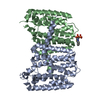
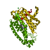
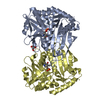
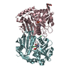
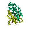
- PDB-5jfq: Geranylgeranyl Pyrophosphate Synthetase from archaeon Geoglobus a... -
+ Open data

- Basic information
| Entry | Database: PDB / ID: 5jfq | ||||||
|---|---|---|---|---|---|---|---|
| Title | Geranylgeranyl Pyrophosphate Synthetase from archaeon Geoglobus acetivorans | ||||||
 Components Components | Geranylgeranyl Pyrophosphate Synthetase | ||||||
 Keywords Keywords |  OXIDOREDUCTASE / Geranylgeranyl Pyrophosphate Synthetase / hyperthermophilic / archaeon OXIDOREDUCTASE / Geranylgeranyl Pyrophosphate Synthetase / hyperthermophilic / archaeon | ||||||
| Function / homology |  Function and homology information Function and homology informationgeranylgeranyl diphosphate biosynthetic process / geranylgeranyl diphosphate synthase / farnesyltranstransferase activity / Transferases; Transferring alkyl or aryl groups, other than methyl groups / geranyl diphosphate biosynthetic process / dimethylallyltranstransferase / farnesyl diphosphate biosynthetic process / (2E,6E)-farnesyl diphosphate synthase / geranyltranstransferase activity / dimethylallyltranstransferase activity / metal ion bindingSimilarity search - Function | ||||||
| Biological species |  Geoglobus acetivorans (archaea) Geoglobus acetivorans (archaea) | ||||||
| Method | X-RAY DIFFRACTION / SYNCHROTRON / MOLECULAR REPLACEMENT / Resolution: 2.51 Å | ||||||
 Authors Authors | Petrova, T. / Boyko, K.M. / Nikolaeva, A.Y. / Stekhanova, T.N. / Mardanov, A.V. / Rakitin, A.L. / Ravin, N.V. / Popov, V.O. | ||||||
 Citation Citation | Journal: Extremophiles / Year: 2018 Title: Structural characterization of geranylgeranyl pyrophosphate synthase GACE1337 from the hyperthermophilic archaeon Geoglobus acetivorans. Authors: Petrova, T.E. / Boyko, K.M. / Nikolaeva, A.Y. / Stekhanova, T.N. / Gruzdev, E.V. / Mardanov, A.V. / Stroilov, V.S. / Littlechild, J.A. / Popov, V.O. / Bezsudnova, E.Y. | ||||||
| History |
|
- Structure visualization
| Structure viewer | Molecule: MolmilJmol/JSmol |
|---|
- Downloads & links
-Download
| PDBx/mmCIF format | 5jfq.cif.gz | 126.7 KB | Display | PDBx/mmCIF format |
|---|---|---|---|---|
| PDB format | pdb5jfq.ent.gz | 97.9 KB | Display | PDB format |
| PDBx/mmJSON format | 5jfq.json.gz | Tree view | PDBx/mmJSON format | |
| Others |  Other downloads Other downloads |
-Validation report
| Arichive directory | https://data.pdbj.org/pub/pdb/validation_reports/jf/5jfqftp://data.pdbj.org/pub/pdb/validation_reports/jf/5jfq | HTTPS FTP |
|---|
-Related structure data
| Related structure data | 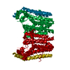 1wy0S S: Starting model for refinement |
|---|---|
| Similar structure data |


-Links
 PDBj
PDBj
- Assembly
| Deposited unit | 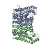
| ||||||||
|---|---|---|---|---|---|---|---|---|---|
| 1 |
| ||||||||
| Unit cell |
|
-Components
| #1: Protein | Mass: 36250.344 Da / Num. of mol.: 2 Source method: isolated from a genetically manipulated source Details: Protein: AIY90378 / Source: (gene. exp.) Geoglobus acetivorans (archaea) / Gene: GACE_1337 / Plasmid: pQE30 / Production host:  Escherichia coli (E. coli) / Strain (production host): DLT1270 / References: UniProt: A0A0A7GEY4 Escherichia coli (E. coli) / Strain (production host): DLT1270 / References: UniProt: A0A0A7GEY4#2: Water | ChemComp-HOH / | Water Mass: 18.015 Da / Num. of mol.: 36 / Source method: isolated from a natural source / Formula: H2O Mass: 18.015 Da / Num. of mol.: 36 / Source method: isolated from a natural source / Formula: H2O |
|---|
-Experimental details
-Experiment
| Experiment | Method: X-RAY DIFFRACTION / Number of used crystals: 1 |
|---|
- Sample preparation
| Crystal | Density Matthews: 2.41 Å3/Da / Density % sol: 48.97 % |
|---|---|
| Crystal grow | Temperature: 310 K / Method: vapor diffusion, hanging drop / pH: 7 Details: 0.1M Tris pH 8.0, 0.1M sodium malonate pH 8.0, 33% PEG 1000 and 10% ethylene glycol mixed with protein in ratio 1:1 over 500 ml of precipitant solution covered with oils |
-Data collection
| Diffraction | Mean temperature: 100 K |
|---|---|
| Diffraction source | Source: SYNCHROTRON / Site: SPring-8  / Beamline: BL41XU / Wavelength: 1 Å / Beamline: BL41XU / Wavelength: 1 Å |
| Detector | Type: RAYONIX MX225HE / Detector: CCD / Date: Jun 15, 2015 |
| Radiation | Protocol: SINGLE WAVELENGTH / Monochromatic (M) / Laue (L): M / Scattering type: x-ray |
| Radiation wavelength | Wavelength: 1 Å / Relative weight: 1 |
| Reflection | Resolution: 2.51→94.08 Å / Num. obs: 24891 / % possible obs: 99.8 % / Redundancy: 19.8 % / Rmerge(I) obs: 0.309 / Net I/σ(I): 6.7 |
| Reflection shell | Resolution: 2.51→2.62 Å / % possible all: 99.7 |
- Processing
| Software |
| |||||||||||||||||||||||||||||||||||||||||||||||||||||||||||||||||||||||||||||||||||||||||||||||||||||||||||||||||||||||
|---|---|---|---|---|---|---|---|---|---|---|---|---|---|---|---|---|---|---|---|---|---|---|---|---|---|---|---|---|---|---|---|---|---|---|---|---|---|---|---|---|---|---|---|---|---|---|---|---|---|---|---|---|---|---|---|---|---|---|---|---|---|---|---|---|---|---|---|---|---|---|---|---|---|---|---|---|---|---|---|---|---|---|---|---|---|---|---|---|---|---|---|---|---|---|---|---|---|---|---|---|---|---|---|---|---|---|---|---|---|---|---|---|---|---|---|---|---|---|---|---|
| Refinement | Method to determine structure: MOLECULAR REPLACEMENT Starting model: 1WY0 Resolution: 2.51→78.112 Å / SU ML: 0.37 / Cross valid method: FREE R-VALUE / σ(F): 0.27 / Phase error: 29 / Stereochemistry target values: ML
| |||||||||||||||||||||||||||||||||||||||||||||||||||||||||||||||||||||||||||||||||||||||||||||||||||||||||||||||||||||||
| Solvent computation | Shrinkage radii: 0.9 Å / VDW probe radii: 1.11 Å / Solvent model: FLAT BULK SOLVENT MODEL | |||||||||||||||||||||||||||||||||||||||||||||||||||||||||||||||||||||||||||||||||||||||||||||||||||||||||||||||||||||||
| Refinement step | Cycle: LAST / Resolution: 2.51→78.112 Å
| |||||||||||||||||||||||||||||||||||||||||||||||||||||||||||||||||||||||||||||||||||||||||||||||||||||||||||||||||||||||
| Refine LS restraints |
| |||||||||||||||||||||||||||||||||||||||||||||||||||||||||||||||||||||||||||||||||||||||||||||||||||||||||||||||||||||||
| LS refinement shell |
|
