 Movie
Movie Controller


+ Open data

- Basic information
| Entry | Database: PDB / ID: 2zom | ||||||
|---|---|---|---|---|---|---|---|
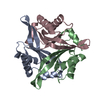
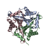
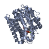
| Title | Crystal structure of CutA1 from Oryza sativa | ||||||
 Components Components | Protein CutA, chloroplast, putative, expressed | ||||||
 Keywords Keywords | UNKNOWN FUNCTION / Trimeric structure /  Protein stability Protein stability | ||||||
| Function / homology |  Function and homology information Function and homology informationresponse to metal ion / protein homotrimerization / chloroplast / copper ion bindingSimilarity search - Function | ||||||
| Biological species |  Oryza sativa subsp. japonica (Japanese rice) Oryza sativa subsp. japonica (Japanese rice) | ||||||
| Method | X-RAY DIFFRACTION / SYNCHROTRON / MOLECULAR REPLACEMENT / Resolution: 3.02 Å | ||||||
 Authors Authors | Kezuka, Y. / Bagautdinov, B. / Katoh, S. / Ohtake, Y. / Yutani, K. / Nonaka, T. / Katoh, E. | ||||||
 Citation Citation | Journal: To be Published Title: Crystal structure of CutA1 from Oryza sativa Authors: Kezuka, Y. / Bagautdinov, B. / Katoh, S. / Ohtake, Y. / Yutani, K. / Nonaka, T. / Katoh, E. #1: Journal: Biochemistry / Year: 2008 Title: Thermodynamic basis for the stabilities of three CutA1s from Pyrococcus horikoshii,Thermus thermophilus, and Oryza sativa, with unusually high denaturation temperatures Authors: Sawano, M. / Yamamoto, H. / Ogasahara, K. / Kidokoro, S. / Katoh, S. / Ohnuma, T. / Katoh, E. / Yokoyama, S. / Yutani, K. | ||||||
| History |
|
- Structure visualization
| Structure viewer | Molecule: MolmilJmol/JSmol |
|---|
- Downloads & links
-Download
| PDBx/mmCIF format | 2zom.cif.gz | 75 KB | Display | PDBx/mmCIF format |
|---|---|---|---|---|
| PDB format | pdb2zom.ent.gz | 57.4 KB | Display | PDB format |
| PDBx/mmJSON format | 2zom.json.gz | Tree view | PDBx/mmJSON format | |
| Others |  Other downloads Other downloads |
-Validation report
| Arichive directory | https://data.pdbj.org/pub/pdb/validation_reports/zo/2zomftp://data.pdbj.org/pub/pdb/validation_reports/zo/2zom | HTTPS FTP |
|---|
-Related structure data
| Related structure data |  2zfhS S: Starting model for refinement |
|---|---|
| Similar structure data |


-Links
 PDBj
PDBj- Assembly
| Deposited unit | 
| ||||||||
|---|---|---|---|---|---|---|---|---|---|
| 1 |
| ||||||||
| Unit cell |
|
-Components
| #1: Protein | Mass: 12538.320 Da / Num. of mol.: 3 / Fragment: UNP residues 65-177 Source method: isolated from a genetically manipulated source Details: Tsunoda et al. (2005). Protein Expression Purif. 42, 268-277. Source: (gene. exp.) Oryza sativa subsp. japonica (Japanese rice)Plasmid: pDEST-his vector / Production host:  Escherichia coli (E. coli) / Strain (production host): BL21(DE3) / References: UniProt: Q109R6 Escherichia coli (E. coli) / Strain (production host): BL21(DE3) / References: UniProt: Q109R6#2: Chemical | Glycerol  Mass: 92.094 Da / Num. of mol.: 3 / Source method: obtained synthetically / Formula: C3H8O3 Mass: 92.094 Da / Num. of mol.: 3 / Source method: obtained synthetically / Formula: C3H8O3#3: Chemical | Sulfate  Mass: 96.063 Da / Num. of mol.: 3 / Source method: obtained synthetically / Formula: SO4 Mass: 96.063 Da / Num. of mol.: 3 / Source method: obtained synthetically / Formula: SO4#4: Water | ChemComp-HOH / | Water Mass: 18.015 Da / Num. of mol.: 3 / Source method: isolated from a natural source / Formula: H2O Mass: 18.015 Da / Num. of mol.: 3 / Source method: isolated from a natural source / Formula: H2O |
|---|
-Experimental details
-Experiment
| Experiment | Method: X-RAY DIFFRACTION / Number of used crystals: 1 |
|---|
- Sample preparation
| Crystal | Density Matthews: 3.28 Å3/Da / Density % sol: 62.47 % |
|---|---|
| Crystal grow | Temperature: 293 K / Method: vapor diffusion, sitting drop / pH: 5.6 Details: 1.8M Ammonium sulfate, 0.09M tri-Sodium citrate dihydrate pH5.6, 0.18M potassium sodium tartrate tetrahydrate, VAPOR DIFFUSION, SITTING DROP, temperature 293K |
-Data collection
| Diffraction | Mean temperature: 100 K |
|---|---|
| Diffraction source | Source: SYNCHROTRON / Site: SPring-8  / Beamline: BL26B1 / Wavelength: 1 Å / Beamline: BL26B1 / Wavelength: 1 Å |
| Detector | Type: RIGAKU JUPITER 210 / Detector: CCD / Date: Jul 4, 2006 / Details: mirrors |
| Radiation | Monochromator: graphite / Protocol: SINGLE WAVELENGTH / Monochromatic (M) / Laue (L): M / Scattering type: x-ray |
| Radiation wavelength | Wavelength: 1 Å / Relative weight: 1 |
| Reflection | Resolution: 3.02→63.628 Å / Num. all: 10138 / Num. obs: 10138 / % possible obs: 100 % / Observed criterion σ(F): 0 / Observed criterion σ(I): 0 / Redundancy: 10.3 % / Biso Wilson estimate: 76.463 Å2 / Rmerge(I) obs: 0.061 / Rsym value: 0.061 / Net I/σ(I): 8 |
| Reflection shell | Resolution: 3.02→3.1 Å / Redundancy: 10.9 % / Rmerge(I) obs: 0.231 / Mean I/σ(I) obs: 3.1 / Num. unique all: 742 / Rsym value: 0.231 / % possible all: 100 |
- Processing
| Software |
| |||||||||||||||||||||||||||||||||||||||||||||||||||||||||||||||||||||||||||||||||||||||||||||||||||||||||||||||||||||||||||||
|---|---|---|---|---|---|---|---|---|---|---|---|---|---|---|---|---|---|---|---|---|---|---|---|---|---|---|---|---|---|---|---|---|---|---|---|---|---|---|---|---|---|---|---|---|---|---|---|---|---|---|---|---|---|---|---|---|---|---|---|---|---|---|---|---|---|---|---|---|---|---|---|---|---|---|---|---|---|---|---|---|---|---|---|---|---|---|---|---|---|---|---|---|---|---|---|---|---|---|---|---|---|---|---|---|---|---|---|---|---|---|---|---|---|---|---|---|---|---|---|---|---|---|---|---|---|---|
| Refinement | Method to determine structure: MOLECULAR REPLACEMENT Starting model: PDB ENTRY 2ZFH Resolution: 3.02→63.61 Å / Cor.coef. Fo:Fc: 0.945 / Cor.coef. Fo:Fc free: 0.881 / SU B: 15.553 / SU ML: 0.283 / Cross valid method: THROUGHOUT / σ(F): 0 / σ(I): 0 / ESU R Free: 0.423 / Stereochemistry target values: MAXIMUM LIKELIHOOD / Details: HYDROGENS HAVE BEEN ADDED IN THE RIDING POSITIONS
| |||||||||||||||||||||||||||||||||||||||||||||||||||||||||||||||||||||||||||||||||||||||||||||||||||||||||||||||||||||||||||||
| Solvent computation | Ion probe radii: 0.8 Å / Shrinkage radii: 0.8 Å / VDW probe radii: 1.2 Å / Solvent model: MASK | |||||||||||||||||||||||||||||||||||||||||||||||||||||||||||||||||||||||||||||||||||||||||||||||||||||||||||||||||||||||||||||
| Displacement parameters | Biso mean: 55.314 Å2
| |||||||||||||||||||||||||||||||||||||||||||||||||||||||||||||||||||||||||||||||||||||||||||||||||||||||||||||||||||||||||||||
| Refinement step | Cycle: LAST / Resolution: 3.02→63.61 Å
| |||||||||||||||||||||||||||||||||||||||||||||||||||||||||||||||||||||||||||||||||||||||||||||||||||||||||||||||||||||||||||||
| Refine LS restraints |
| |||||||||||||||||||||||||||||||||||||||||||||||||||||||||||||||||||||||||||||||||||||||||||||||||||||||||||||||||||||||||||||
| LS refinement shell | Resolution: 3.02→3.098 Å / Total num. of bins used: 20
|
