 Movie
Movie Controller


+ Open data

- Basic information
| Entry | Database: PDB / ID: 1osc | ||||||
|---|---|---|---|---|---|---|---|
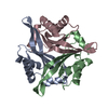
| Title | Crystal structure of rat CUTA1 at 2.15 A resolution | ||||||
 Components Components | similar to divalent cation tolerant protein CUTA | ||||||
 Keywords Keywords | UNKNOWN FUNCTION / CUTA / copper resistance /  Structural Proteomics in Europe / SPINE / Structural Genomics Structural Proteomics in Europe / SPINE / Structural Genomics | ||||||
| Function / homology |  Function and homology information Function and homology informationresponse to metal ion / protein localization / copper ion binding / enzyme binding / membraneSimilarity search - Function | ||||||
| Biological species |  Rattus norvegicus (Norway rat) Rattus norvegicus (Norway rat) | ||||||
| Method | X-RAY DIFFRACTION / SYNCHROTRON / MOLECULAR REPLACEMENT / Resolution: 2.15 Å | ||||||
 Authors Authors | Arnesano, F. / Banci, L. / Benvenuti, M. / Bertini, I. / Calderone, V. / Mangani, S. / Viezzoli, M.S. / Structural Proteomics in Europe (SPINE) | ||||||
 Citation Citation | Journal: J.Biol.Chem. / Year: 2003 Title: The Evolutionarily Conserved Trimeric Structure of CutA1 Proteins Suggests a Role in Signal Transduction Authors: Arnesano, F. / Banci, L. / Benvenuti, M. / Bertini, I. / Calderone, V. / Mangani, S. / Viezzoli, M.S. #1: Journal: To be PublishedTitle: Structure of protein TM1056, CUTA Authors: SAVCHENKO, A. / ZHANG, R. / JOACHIMIAK, A. / EDWARDS, A. / AKARINA, T. #2: Journal: To be PublishedTitle: The trimeric structure of CutA1 proteins of bacteria and mammals is reminiscent of the architecture of signal transduction proteins. Authors: Calderone, V. / Mangani, S. / Benvenuti, M. / Viezzoli, M.S. / Banci, L. / Bertini, I. | ||||||
| History |
|
- Structure visualization
| Structure viewer | Molecule: MolmilJmol/JSmol |
|---|
- Downloads & links
-Download
| PDBx/mmCIF format | 1osc.cif.gz | 147.7 KB | Display | PDBx/mmCIF format |
|---|---|---|---|---|
| PDB format | pdb1osc.ent.gz | 117.2 KB | Display | PDB format |
| PDBx/mmJSON format | 1osc.json.gz | Tree view | PDBx/mmJSON format | |
| Others |  Other downloads Other downloads |
-Validation report
| Arichive directory | https://data.pdbj.org/pub/pdb/validation_reports/os/1oscftp://data.pdbj.org/pub/pdb/validation_reports/os/1osc | HTTPS FTP |
|---|
-Related structure data
| Related structure data |  1naqSC S: Starting model for refinement C: citing same article ( |
|---|---|
| Similar structure data | |
| Other databases |


-Links
 PDBj
PDBj- Assembly
| Deposited unit | 
| ||||||||||||||||||||||||||||||||||||||||||||||||||||||||||||||||||||||||||||||||
|---|---|---|---|---|---|---|---|---|---|---|---|---|---|---|---|---|---|---|---|---|---|---|---|---|---|---|---|---|---|---|---|---|---|---|---|---|---|---|---|---|---|---|---|---|---|---|---|---|---|---|---|---|---|---|---|---|---|---|---|---|---|---|---|---|---|---|---|---|---|---|---|---|---|---|---|---|---|---|---|---|---|
| 1 | 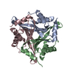
| ||||||||||||||||||||||||||||||||||||||||||||||||||||||||||||||||||||||||||||||||
| 2 | 
| ||||||||||||||||||||||||||||||||||||||||||||||||||||||||||||||||||||||||||||||||
| 3 |
| ||||||||||||||||||||||||||||||||||||||||||||||||||||||||||||||||||||||||||||||||
| Unit cell |
| ||||||||||||||||||||||||||||||||||||||||||||||||||||||||||||||||||||||||||||||||
| Noncrystallographic symmetry (NCS) | NCS domain:
NCS domain segments: Component-ID: 1 / End auth comp-ID: VAL / End label comp-ID: VAL / Refine code: 5
NCS ensembles :
| ||||||||||||||||||||||||||||||||||||||||||||||||||||||||||||||||||||||||||||||||
| Details | The biological functional unit is a trimer; the asymetric unit is made of two trimers |
-Components
| #1: Protein | Mass: 13600.518 Da / Num. of mol.: 6 / Fragment: residue 44-169 Source method: isolated from a genetically manipulated source Source: (gene. exp.) Rattus norvegicus (Norway rat) / Gene: CutA, CutA1 / Production host:  Escherichia coli (E. coli) / References: UniProt: Q6MGD0 Escherichia coli (E. coli) / References: UniProt: Q6MGD0#2: Water | ChemComp-HOH / | Water Mass: 18.015 Da / Num. of mol.: 405 / Source method: isolated from a natural source / Formula: H2O Mass: 18.015 Da / Num. of mol.: 405 / Source method: isolated from a natural source / Formula: H2O |
|---|
-Experimental details
-Experiment
| Experiment | Method: X-RAY DIFFRACTION / Number of used crystals: 1 |
|---|
- Sample preparation
| Crystal | Density Matthews: 2.53 Å3/Da / Density % sol: 51.04 % | ||||||||||||||||||||||||||||||||||||||||||
|---|---|---|---|---|---|---|---|---|---|---|---|---|---|---|---|---|---|---|---|---|---|---|---|---|---|---|---|---|---|---|---|---|---|---|---|---|---|---|---|---|---|---|---|
| Crystal grow | Temperature: 298 K / Method: vapor diffusion, sitting drop / pH: 4.6 Details: Na Acetate, CuSO4, CaCl2, pH 4.6, VAPOR DIFFUSION, SITTING DROP, temperature 298K | ||||||||||||||||||||||||||||||||||||||||||
| Crystal grow | *PLUS Temperature: 20 ℃ / Method: vapor diffusion | ||||||||||||||||||||||||||||||||||||||||||
| Components of the solutions | *PLUS
|
-Data collection
| Diffraction | Mean temperature: 100 K |
|---|---|
| Diffraction source | Source: SYNCHROTRON / Site: ESRF  / Beamline: ID14-1 / Wavelength: 0.9322 Å / Beamline: ID14-1 / Wavelength: 0.9322 Å |
| Detector | Type: ADSC QUANTUM 4 / Detector: CCD / Date: Feb 1, 2003 / Details: Diamond (111), Ge(220) |
| Radiation | Monochromator: Diamond (111), Ge(220) / Protocol: SINGLE WAVELENGTH / Monochromatic (M) / Laue (L): M / Scattering type: x-ray |
| Radiation wavelength | Wavelength: 0.9322 Å / Relative weight: 1 |
| Reflection | Resolution: 2.15→20 Å / Num. all: 43312 / Num. obs: 43312 / Observed criterion σ(F): 0 / Observed criterion σ(I): 0 / Redundancy: 6.5 % / Rmerge(I) obs: 0.073 / Rsym value: 0.073 / Net I/σ(I): 5.3 |
| Reflection shell | Resolution: 2.15→2.2 Å / Redundancy: 4.8 % / Rmerge(I) obs: 0.47 / Mean I/σ(I) obs: 1.8 / Rsym value: 0.47 / % possible all: 99.9 |
| Reflection | *PLUS Lowest resolution: 40 Å / Num. obs: 46060 / % possible obs: 100 % / Num. measured all: 300781 |
| Reflection shell | *PLUS % possible obs: 100 % / Num. unique obs: 6646 / Num. measured obs: 32115 / Rmerge(I) obs: 0.47 / Mean I/σ(I) obs: 1.7 |
- Processing
| Software |
| |||||||||||||||||||||||||||||||||||||||||||||||||||||||||||||||||||||||||||||||||||||||||||||||||||||||||||||||||||||||||||||||||||||||||||||||||||||||||||||||||||||||||||||||
|---|---|---|---|---|---|---|---|---|---|---|---|---|---|---|---|---|---|---|---|---|---|---|---|---|---|---|---|---|---|---|---|---|---|---|---|---|---|---|---|---|---|---|---|---|---|---|---|---|---|---|---|---|---|---|---|---|---|---|---|---|---|---|---|---|---|---|---|---|---|---|---|---|---|---|---|---|---|---|---|---|---|---|---|---|---|---|---|---|---|---|---|---|---|---|---|---|---|---|---|---|---|---|---|---|---|---|---|---|---|---|---|---|---|---|---|---|---|---|---|---|---|---|---|---|---|---|---|---|---|---|---|---|---|---|---|---|---|---|---|---|---|---|---|---|---|---|---|---|---|---|---|---|---|---|---|---|---|---|---|---|---|---|---|---|---|---|---|---|---|---|---|---|---|---|---|---|
| Refinement | Method to determine structure: MOLECULAR REPLACEMENT Starting model: 1NAQ Resolution: 2.15→20 Å / Cor.coef. Fo:Fc: 0.945 / Cor.coef. Fo:Fc free: 0.907 / SU B: 6.271 / SU ML: 0.161 / Isotropic thermal model: isotropic / Cross valid method: THROUGHOUT / ESU R: 0.236 / ESU R Free: 0.215 / Stereochemistry target values: MAXIMUM LIKELIHOOD
| |||||||||||||||||||||||||||||||||||||||||||||||||||||||||||||||||||||||||||||||||||||||||||||||||||||||||||||||||||||||||||||||||||||||||||||||||||||||||||||||||||||||||||||||
| Solvent computation | Ion probe radii: 0.8 Å / Shrinkage radii: 0.8 Å / VDW probe radii: 1.4 Å / Solvent model: BABINET MODEL WITH MASK | |||||||||||||||||||||||||||||||||||||||||||||||||||||||||||||||||||||||||||||||||||||||||||||||||||||||||||||||||||||||||||||||||||||||||||||||||||||||||||||||||||||||||||||||
| Displacement parameters | Biso mean: 57.5 Å2
| |||||||||||||||||||||||||||||||||||||||||||||||||||||||||||||||||||||||||||||||||||||||||||||||||||||||||||||||||||||||||||||||||||||||||||||||||||||||||||||||||||||||||||||||
| Refinement step | Cycle: LAST / Resolution: 2.15→20 Å
| |||||||||||||||||||||||||||||||||||||||||||||||||||||||||||||||||||||||||||||||||||||||||||||||||||||||||||||||||||||||||||||||||||||||||||||||||||||||||||||||||||||||||||||||
| Refine LS restraints |
| |||||||||||||||||||||||||||||||||||||||||||||||||||||||||||||||||||||||||||||||||||||||||||||||||||||||||||||||||||||||||||||||||||||||||||||||||||||||||||||||||||||||||||||||
| Refine LS restraints NCS | Refine-ID: X-RAY DIFFRACTION
| |||||||||||||||||||||||||||||||||||||||||||||||||||||||||||||||||||||||||||||||||||||||||||||||||||||||||||||||||||||||||||||||||||||||||||||||||||||||||||||||||||||||||||||||
| LS refinement shell | Resolution: 2.15→2.205 Å / Total num. of bins used: 20 /
| |||||||||||||||||||||||||||||||||||||||||||||||||||||||||||||||||||||||||||||||||||||||||||||||||||||||||||||||||||||||||||||||||||||||||||||||||||||||||||||||||||||||||||||||
| Software | *PLUS Version: 5.1.24 / Classification: refinement | |||||||||||||||||||||||||||||||||||||||||||||||||||||||||||||||||||||||||||||||||||||||||||||||||||||||||||||||||||||||||||||||||||||||||||||||||||||||||||||||||||||||||||||||
| Refinement | *PLUS Lowest resolution: 20 Å / Rfactor Rfree: 0.26 / Rfactor Rwork: 0.189 | |||||||||||||||||||||||||||||||||||||||||||||||||||||||||||||||||||||||||||||||||||||||||||||||||||||||||||||||||||||||||||||||||||||||||||||||||||||||||||||||||||||||||||||||
| Solvent computation | *PLUS | |||||||||||||||||||||||||||||||||||||||||||||||||||||||||||||||||||||||||||||||||||||||||||||||||||||||||||||||||||||||||||||||||||||||||||||||||||||||||||||||||||||||||||||||
| Displacement parameters | *PLUS | |||||||||||||||||||||||||||||||||||||||||||||||||||||||||||||||||||||||||||||||||||||||||||||||||||||||||||||||||||||||||||||||||||||||||||||||||||||||||||||||||||||||||||||||
| Refine LS restraints | *PLUS
|
