 Movie
Movie Controller


+ Open data

- Basic information
| Entry | Database: PDB / ID: 2aem | ||||||
|---|---|---|---|---|---|---|---|
| Title | Crystal Structures of the MthK RCK Domain | ||||||
 Components Components | Calcium-gated potassium channel mthK | ||||||
 Keywords Keywords | METAL TRANSPORT /  MEMBRANE PROTEIN / Rossmann fold / helix-turn-helix MEMBRANE PROTEIN / Rossmann fold / helix-turn-helix | ||||||
| Function / homology |  Function and homology information Function and homology informationmonoatomic cation transmembrane transporter activity / potassium ion transport / identical protein binding / metal ion binding / plasma membraneSimilarity search - Function | ||||||
| Biological species |   Methanothermobacter thermautotrophicus (archaea) Methanothermobacter thermautotrophicus (archaea) | ||||||
| Method | X-RAY DIFFRACTION / SYNCHROTRON / MOLECULAR REPLACEMENT / Resolution: 2.8 Å | ||||||
 Authors Authors | Dong, J. / Shi, N. / Berke, I. / Chen, L. / Jiang, Y. | ||||||
 Citation Citation | Journal: J.Biol.Chem. / Year: 2005 Title: Structures of the MthK RCK Domain and the Effect of Ca2+ on Gating Ring Stability Authors: Dong, J. / Shi, N. / Berke, I. / Chen, L. / Jiang, Y. #1: Journal: Nature / Year: 2002Title: Crystal structure and mechanism of a calcium-gated potassium channel Authors: Jiang, Y. / Lee, A. / Chen, J. / Cadene, M. / Chait, B.T. / MacKinnon, R. | ||||||
| History |
|
- Structure visualization
| Structure viewer | Molecule: MolmilJmol/JSmol |
|---|
- Downloads & links
-Download
| PDBx/mmCIF format | 2aem.cif.gz | 55.8 KB | Display | PDBx/mmCIF format |
|---|---|---|---|---|
| PDB format | pdb2aem.ent.gz | 40.2 KB | Display | PDB format |
| PDBx/mmJSON format | 2aem.json.gz | Tree view | PDBx/mmJSON format | |
| Others |  Other downloads Other downloads |
-Validation report
| Arichive directory | https://data.pdbj.org/pub/pdb/validation_reports/ae/2aemftp://data.pdbj.org/pub/pdb/validation_reports/ae/2aem | HTTPS FTP |
|---|
-Related structure data
| Related structure data |  2aefC  2aejC  1lnqS S: Starting model for refinement C: citing same article ( |
|---|---|
| Similar structure data |


-Links
 PDBj
PDBj

- Assembly
| Deposited unit | 
| ||||||||
|---|---|---|---|---|---|---|---|---|---|
| 1 | 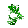
| ||||||||
| Unit cell |
| ||||||||
| Components on special symmetry positions |
|
-Components
| #1: Protein | Mass: 25914.584 Da / Num. of mol.: 1 Source method: isolated from a genetically manipulated source Source: (gene. exp.) Methanothermobacter thermautotrophicus (archaea)Gene: mthK / Plasmid: pQE70 / Production host:  Escherichia coli (E. coli) / Strain (production host): M15 / References: UniProt: O27564 Escherichia coli (E. coli) / Strain (production host): M15 / References: UniProt: O27564 |
|---|---|
| #2: Water | ChemComp-HOH / Water Mass: 18.015 Da / Num. of mol.: 5 / Source method: isolated from a natural source / Formula: H2O Mass: 18.015 Da / Num. of mol.: 5 / Source method: isolated from a natural source / Formula: H2O |
-Experimental details
-Experiment
| Experiment | Method: X-RAY DIFFRACTION / Number of used crystals: 1 |
|---|
- Sample preparation
| Crystal | Density Matthews: 4.17 Å3/Da / Density % sol: 70 % |
|---|---|
| Crystal grow | Temperature: 293 K / Method: vapor diffusion, sitting drop / pH: 5.5 Details: PEG 400, Ammonium sulfate, cacodylate , pH 5.5, VAPOR DIFFUSION, SITTING DROP, temperature 293K |
-Data collection
| Diffraction | Mean temperature: 100 K |
|---|---|
| Diffraction source | Source: SYNCHROTRON / Site: APS  / Beamline: 19-BM / Wavelength: 0.979 Å / Beamline: 19-BM / Wavelength: 0.979 Å |
| Detector | Type: ADSC QUANTUM 4 / Detector: CCD / Date: Jun 5, 2004 |
| Radiation | Protocol: SINGLE WAVELENGTH / Monochromatic (M) / Laue (L): M / Scattering type: x-ray |
| Radiation wavelength | Wavelength: 0.979 Å / Relative weight: 1 |
| Reflection | Resolution: 2.8→35.8 Å / Num. all: 10766 / Num. obs: 10728 / % possible obs: 99.7 % / Observed criterion σ(I): 2.77 / Redundancy: 6.1 % / Rmerge(I) obs: 0.068 / Rsym value: 0.068 / Net I/σ(I): 23 |
| Reflection shell | Resolution: 2.8→2.9 Å / Redundancy: 4.1 % / Rmerge(I) obs: 0.524 / Num. unique all: 1068 / Rsym value: 0.524 / % possible all: 100 |
- Processing
| Software |
| |||||||||||||||||||||||||
|---|---|---|---|---|---|---|---|---|---|---|---|---|---|---|---|---|---|---|---|---|---|---|---|---|---|---|
| Refinement | Method to determine structure: MOLECULAR REPLACEMENT Starting model: pdb entry 1LNQ Resolution: 2.8→35.8 Å / σ(F): 0 / σ(I): 0 / Stereochemistry target values: Engh & Huber
| |||||||||||||||||||||||||
| Refinement step | Cycle: LAST / Resolution: 2.8→35.8 Å
|
