Movie
Movie Controller
 Structure viewers
 About EMN search


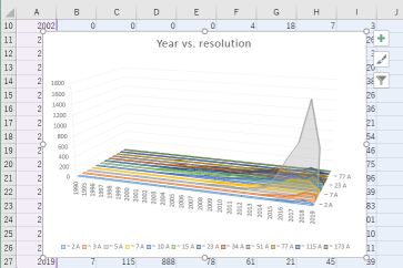
-Search query
-Search result
Showing 1 - 50 of 124 items for (author: lei & ht)

EMDB-40180: 
MsbA bound to cerastecin C
Method: single particle / : Chen Y, Klein D

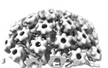
EMDB-18482:
Herpes simplex virus 1 capsid (WT) vertices in perinuclear NEC-coated vesicles determined in situ
Method: subtomogram averaging / : Mironova Y, Prazak V, Vasishtan D
EMDB-18484:
Herpes simplex virus 1 nuclear egress complex (WT) determined in situ from perinuclear vesicles
Method: subtomogram averaging / : Mironova Y, Prazak V, Vasishtan D

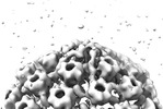
EMDB-17974:
Pseudorabies virus cytosolic C-capsid (US3 KO) vertices determined in situ
Method: subtomogram averaging / : Prazak V, Grange M, Vasishtan D

EMDB-17975:
Pseudorabies virus primary enveloped (perinuclear) C-capsid (US3 KO) vertices determined in situ
Method: subtomogram averaging / : Prazak V, Grange M, Vasishtan D

EMDB-17976:
Pseudorabies nuclear C-capsids (US3 KO) vertices determined in situ
Method: subtomogram averaging / : Prazak V, Grange M, Vasishtan D
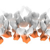
EMDB-18473:
Subtomogram average of pseudorabies virus nuclear egress complex helical form (UL31/34) determined in situ
Method: subtomogram averaging / : Prazak V, Grange M, Vasishtan D
EMDB-18474:
Subtomogram average of pseudorabies virus nuclear egress complex (UL31/34) determined in situ
Method: subtomogram averaging / : Prazak V, Grange M, Vasishtan D

EMDB-18479:
Pseudorabies virus cytosolic C-capsid (WT) vertices determined in situ
Method: subtomogram averaging / : Mironova Y, Prazak V, Vasishtan D

EMDB-18480:
Pseudorabies virus nuclear C-capsid (WT) vertices determined in situ
Method: subtomogram averaging / : Mironova Y, Prazak V, Vasishtan D
EMDB-18481:
Herpes simplex virus 1 cytosolic C-capsid (WT) vertices determined in situ
Method: subtomogram averaging / : Mironova Y, Prazak V, Vasishtan D
EMDB-18483:
Herpes simplex virus 1 nuclear C-capsid (WT) vertices determined in situ
Method: subtomogram averaging / : Mironova Y, Prazak V, Vasishtan D

EMDB-27641:
The structure of the interleukin 11 signalling complex, truncated gp130
Method: single particle / : Metcalfe RD, Hanssen E, Griffin MDW

EMDB-27642:
The structure of the IL-11 signalling complex, with full-length extracellular gp130
Method: single particle / : Metcalfe RD, Hanssen E, Griffin MDW

PDB-8dps:
The structure of the interleukin 11 signalling complex, truncated gp130
Method: single particle / : Metcalfe RD, Hanssen E, Griffin MDW

PDB-8dpt:
The structure of the IL-11 signalling complex, with full-length extracellular gp130
Method: single particle / : Metcalfe RD, Hanssen E, Griffin MDW
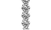
EMDB-16458:
Electron cryo-tomography and subtomogram averaging of cytoplasmic lattice filaments from mammalian oocytes
Method: subtomogram averaging / : Petrovic A, Bauerlein FJB, Jentoft IMA, Schuh M, Fernandez-Busnadiego R

EMDB-16472:
In situ cryo-electron tomogram of cytoplasmic lattice filaments from a mouse oocyte
Method: electron tomography / : Bauerlein FJB, Jentoft IMA, Petrovic A, Fernandez-Busnadiego R, Schuh M

EMDB-41158:
CS2it1p2_F7K local refinement for HCMV Trimer in complex with CS2it1p2_F7K Fab and CS4tt1p1_E3K Fab
Method: single particle / : Goldsmith JA, McLellan JS

EMDB-41180:
Global reconstruction for HCMV Pentamer in complex with CS2pt1p2_A10L Fab and CS3pt1p4_C1L Fab
Method: single particle / : Goldsmith JG, McLellan JS

EMDB-34813:
Structure of transforming growth factor beta induced protein (TGFBIp) G623R fibril
Method: helical / : Low JYK, Pervushin K

PDB-8hia:
Structure of transforming growth factor beta induced protein (TGFBIp) G623R fibril
Method: helical / : Low JYK, Pervushin K
EMDB-33845:
Cryo-EM map of Rpd3S complex
Method: single particle / : Li HT, Yan CY, Guan HP, Wang P
EMDB-33846:
Cryo-EM map of Rpd3S:head-bridge-right arm
Method: single particle / : Li HT, Yan CY, Guan HP, Wang P
EMDB-33847:
Cryo-EM map of Rpd3S:bridge-left arm
Method: single particle / : Li HT, Yan CY, Guan HP, Wang P

EMDB-33848:
Cryo-EM map of Eaf3 CHD bound to H3K36me3 nucleosome
Method: single particle / : Li HT, Yan CY, Guan HP, Wang P

EMDB-33849:
Cryo-EM map of Rpd3S in loose-state Rpd3S-NCP complex
Method: single particle / : Li HT, Yan CY, Guan HP, Wang P

EMDB-33850:
Cryo-EM map of Rpd3S in close-state Rpd3S-NCP complex
Method: single particle / : Li HT, Yan CY, Guan HP, Wang P

EMDB-33851:
Cryo-EM map of Rpd3S complex bound to H3K36me3 nucleosome in close state
Method: single particle / : Li HT, Yan CY, Guan HP, Wang P

EMDB-33852:
Cryo-EM msp of Rpd3S complex bound to H3K36me3 nucleosome in loose state
Method: single particle / : Li HT, Yan CY, Guan HP, Wang P
EMDB-27148:
Zebrafish MFSD2A isoform B in inward open ligand bound conformation
Method: single particle / : Nguyen C, Lei HT, Lai LTF, Gallentino MJ, Mu X, Matthies D, Gonen T
EMDB-27149:
Zebrafish MFSD2A isoform B in inward open ligand-free conformation
Method: single particle / : Nguyen C, Lei HT, Lai LTF, Gallentino MJ, Mu X, Matthies D, Gonen T
EMDB-27150:
Zebrafish MFSD2A isoform B in inward open ligand 1A conformation
Method: single particle / : Nguyen C, Lei HT, Lai LTF, Gallentino MJ, Mu X, Matthies D, Gonen T
EMDB-27151:
Zebrafish MFSD2A isoform B in inward open ligand 1B conformation
Method: single particle / : Nguyen C, Lei HT, Lai LTF, Gallentino MJ, Mu X, Matthies D, Gonen T
EMDB-27152:
Zebrafish MFSD2A isoform B in inward open ligand 2B conformation
Method: single particle / : Nguyen C, Lei HT, Lai LTF, Gallentino MJ, Mu X, Matthies D, Gonen T
EMDB-27153:
Zebrafish MFSD2A isoform B in inward open ligand 3C conformation
Method: single particle / : Nguyen C, Lei HT, Lai LTF, Gallentino MJ, Mu X, Matthies D, Gonen T
EMDB-16128:
Tomogram of a late endosome of A549 cell infected with influenza A virus.
Method: electron tomography / : Klein S, Chlanda P

EMDB-16129:
Tomogram of a late endosome of A549 cell infected with influenza A virus (Figure 6C).
Method: electron tomography / : Klein S, Chlanda P

EMDB-16130:
Tomogram of a late endosome of A549 cell infected with influenza A virus (Figure 6E)
Method: electron tomography / : Klein S, Chlanda P
EMDB-16131:
Tomogram of a late endosome of A549 cell infected with influenza A virus (Figure 6G)
Method: electron tomography / : Klein S, Chlanda P

EMDB-16132:
Tomogram of a late endosome of A549 cell infected with influenza A virus (Figure 6I,K,M)
Method: electron tomography / : Klein S, Chlanda P
EMDB-16133:
Tomogram of a late endosome of A549 cell infected with influenza A virus (Figure 6O)
Method: electron tomography / : Klein S, Chlanda P
EMDB-28092:
Negative stain EM map of SARS-CoV-2 Spike in complex with CoVIC-093
Method: single particle / : Callaway H, Li H, Yu X, Shek J, Saphire EO
EMDB-28090:
Negative stain EM map of SARS-CoV-2 Spike in complex with CoVIC-040
Method: single particle / : Li H, Callaway H, Yu X, Shek J, Saphire EO
EMDB-28091:
Negative stain EM map of SARS-CoV-2 Spike in complex with CoVIC-045
Method: single particle / : Li H, Callaway H, Yu X, Shek J, Saphire EO
EMDB-28093:
Negative stain EM map of SARS-CoV-2 Spike in complex with CoVIC-156
Method: single particle / : Shek J, Callaway H, Li H, Yu X, Saphire EO

EMDB-27243:
Cryo-EM map of MORF-WHs in complex with 197bp nucleosome aided by scFv
Method: single particle / : Zhou BR
EMDB-28094:
Negative stain EM map of SARS-CoV-2 Spike in complex with CoVIC-234
Method: single particle / : Callaway H, Li H, Yu X, Shek J, Saphire EO
EMDB-28095:
Negative stain EM map of SARS-CoV-2 Spike in complex with CoVIC-260
Method: single particle / : Callaway H, Li H, Yu X, Shek J, Saphire EO
Pages:
 wwPDB to switch to version 3 of the EMDB data model