 Movie
Movie Controller


+ Open data

- Basic information
| Entry | Database: PDB / ID: 8v24 | |||||||||
|---|---|---|---|---|---|---|---|---|---|---|
| Title | LapB cytoplasmic domain in complex with LpxC | |||||||||
 Components Components |
| |||||||||
 Keywords Keywords |  PROTEIN BINDING / adaptor / complex / deacetylase / LPS / LpxC / LapB(YciM) PROTEIN BINDING / adaptor / complex / deacetylase / LPS / LpxC / LapB(YciM) | |||||||||
| Function / homology |  Function and homology information Function and homology informationlipopolysaccharide metabolic process / UDP-3-O-acyl-N-acetylglucosamine deacetylase / UDP-3-O-[3-hydroxymyristoyl] N-acetylglucosamine deacetylase activity / UDP-3-O-acyl-N-acetylglucosamine deacetylase activity / regulation of lipid biosynthetic process / lipid A biosynthetic process / cytoplasmic side of plasma membrane / iron ion binding / metal ion bindingSimilarity search - Function | |||||||||
| Biological species |  Escherichia coli CFT073 (bacteria) Escherichia coli CFT073 (bacteria) | |||||||||
| Method | ELECTRON MICROSCOPY / single particle reconstruction / cryo EM / Resolution: 3.6 Å | |||||||||
 Authors Authors | Mi, W. / Shu, S. | |||||||||
| Funding support |  United States, 2items United States, 2items
| |||||||||
 Citation Citation | Journal: Proc Natl Acad Sci U S A / Year: 2024 Title: Dual function of LapB (YciM) in regulating lipopolysaccharide synthesis. Authors: Sheng Shu / Yuko Tsutsui / Rajkanwar Nathawat / Wei Mi / Abstract: Levels of lipopolysaccharide (LPS), an essential glycolipid on the surface of most gram-negative bacteria, are tightly controlled-making LPS synthesis a promising target for developing new ...Levels of lipopolysaccharide (LPS), an essential glycolipid on the surface of most gram-negative bacteria, are tightly controlled-making LPS synthesis a promising target for developing new antibiotics. adaptor protein LapB (YciM) plays an important role in regulating LPS synthesis by promoting degradation of LpxC, a deacetylase that catalyzes the first committed step in LPS synthesis. Under conditions where LPS is abundant, LapB recruits LpxC to the AAA+ protease FtsH for degradation. LapB achieves this by simultaneously interacting with FtsH through its transmembrane helix and LpxC through its cytoplasmic domain. Here, we describe a cryo-EM structure of the complex formed between LpxC and the cytoplasmic domain of LapB (LapB). The structure reveals how LapB exploits both its tetratricopeptide repeat (TPR) motifs and rubredoxin domain to interact with LpxC. Through both in vitro and in vivo analysis, we show that mutations at the LapB/LpxC interface prevent LpxC degradation. Unexpectedly, binding to LapB also inhibits the enzymatic activity of LpxC through allosteric effects reminiscent of LpxC activation by MurA in Our findings argue that LapB regulates LPS synthesis in two steps: In the first step, LapB inhibits the activity of LpxC, and in the second step, it commits LpxC to degradation by FtsH. | |||||||||
| History |
|
- Structure visualization
| Structure viewer | Molecule: MolmilJmol/JSmol |
|---|
- Downloads & links
-Download
| PDBx/mmCIF format | 8v24.cif.gz | 415.8 KB | Display | PDBx/mmCIF format |
|---|---|---|---|---|
| PDB format | pdb8v24.ent.gz | 343 KB | Display | PDB format |
| PDBx/mmJSON format | 8v24.json.gz | Tree view | PDBx/mmJSON format | |
| Others |  Other downloads Other downloads |
-Validation report
| Arichive directory | https://data.pdbj.org/pub/pdb/validation_reports/v2/8v24ftp://data.pdbj.org/pub/pdb/validation_reports/v2/8v24 | HTTPS FTP |
|---|
-Related structure data
| Related structure data | 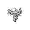 42897MC M: map data used to model this data C: citing same article ( |
|---|---|
| Similar structure data |
-Links
 PDBj
PDBj


- Assembly
| Deposited unit | 
|
|---|---|
| 1 |
|
-Components
| #1: Protein | Mass: 44588.969 Da / Num. of mol.: 2 Source method: isolated from a genetically manipulated source Source: (gene. exp.) Escherichia coli CFT073 (bacteria) / Strain: CFT073 / ATCC 700928 / UPEC / Gene: lapB / Production host: Escherichia coli K-12 (bacteria) / References: UniProt: P0AB59#2: Protein | Mass: 33995.871 Da / Num. of mol.: 2 Source method: isolated from a genetically manipulated source Source: (gene. exp.) Escherichia coli CFT073 (bacteria) / Strain: CFT073 / ATCC 700928 / UPEC / Gene: lpxC / Production host: Escherichia coli K-12 (bacteria) / References: UniProt: P0A726#3: Chemical | ChemComp-ZN /   Mass: 65.409 Da / Num. of mol.: 4 / Source method: obtained synthetically / Formula: Zn / Feature type: SUBJECT OF INVESTIGATION Mass: 65.409 Da / Num. of mol.: 4 / Source method: obtained synthetically / Formula: Zn / Feature type: SUBJECT OF INVESTIGATION#4: Chemical |   Mass: 791.672 Da / Num. of mol.: 2 / Source method: obtained synthetically / Formula: C29H51N3O18P2 / Feature type: SUBJECT OF INVESTIGATION Mass: 791.672 Da / Num. of mol.: 2 / Source method: obtained synthetically / Formula: C29H51N3O18P2 / Feature type: SUBJECT OF INVESTIGATION#5: Chemical | Acetate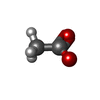  Mass: 59.044 Da / Num. of mol.: 2 / Source method: obtained synthetically / Formula: C2H3O2 / Feature type: SUBJECT OF INVESTIGATION Mass: 59.044 Da / Num. of mol.: 2 / Source method: obtained synthetically / Formula: C2H3O2 / Feature type: SUBJECT OF INVESTIGATIONHas ligand of interest | Y | |
|---|
-Experimental details
-Experiment
| Experiment | Method: ELECTRON MICROSCOPY |
|---|---|
| EM experiment | Aggregation state: PARTICLE / 3D reconstruction method: single particle reconstruction |
- Sample preparation
| Component | Name: LapB/LpxC / Type: COMPLEX / Entity ID: #1-#2 / Source: RECOMBINANT |
|---|---|
| Molecular weight | Experimental value: NO |
| Source (natural) | Organism: Escherichia coli CFT073 (bacteria) |
| Source (recombinant) | Organism: Escherichia coli K-12 (bacteria) |
| Buffer solution | pH: 7.8 |
| Specimen | Conc.: 10 mg/ml / Embedding applied: NO / Shadowing applied: NO / Staining applied: NO / Vitrification applied: YES |
| Specimen support | Grid material: GOLD / Grid mesh size: 300 divisions/in. / Grid type: C-flat-1.2/1.3 |
| Vitrification | Instrument: GATAN CRYOPLUNGE 3 / Cryogen name: ETHANE |
- Electron microscopy imaging
| Experimental equipment |  Model: Titan Krios / Image courtesy: FEI Company |
|---|---|
| Microscopy | Model: FEI TITAN KRIOS |
| Electron gun | Electron source: FIELD EMISSION GUN / Accelerating voltage: 300 kV / Illumination mode: FLOOD BEAM |
| Electron lens | Mode: BRIGHT FIELDBright-field microscopy / Nominal magnification: 81000 X / Nominal defocus max: 2500 nm / Nominal defocus min: 1000 nm / Cs: 2.7 mm / C2 aperture diameter: 100 µm |
| Image recording | Average exposure time: 2.6 sec. / Electron dose: 50 e/Å2 / Film or detector model: GATAN K3 (6k x 4k) / Num. of grids imaged: 1 |
| EM imaging optics | Energyfilter name: GIF Quantum LS / Energyfilter slit width: 20 eV |
- Processing
| EM software |
| ||||||||||||||||||||||||||||||||||||||||
|---|---|---|---|---|---|---|---|---|---|---|---|---|---|---|---|---|---|---|---|---|---|---|---|---|---|---|---|---|---|---|---|---|---|---|---|---|---|---|---|---|---|
| CTF correction | Type: PHASE FLIPPING AND AMPLITUDE CORRECTION | ||||||||||||||||||||||||||||||||||||||||
| 3D reconstruction | Resolution: 3.6 Å / Resolution method: FSC 0.143 CUT-OFF / Num. of particles: 1171191 / Num. of class averages: 1 / Symmetry type: POINT | ||||||||||||||||||||||||||||||||||||||||
| Refine LS restraints |
|
