lipopolysaccharide metabolic process / UDP-3-O-acyl-N-acetylglucosamine deacetylase / UDP-3-O-acyl-N-acetylglucosamine deacetylase activity / regulation of lipid biosynthetic process / lipid A biosynthetic process / cytoplasmic side of plasma membrane / iron ion binding / metal ion binding / membrane Similarity search - Function
National Institutes of Health/National Institute of General Medical Sciences (NIH/NIGMS)
R01GM137068
United States
National Institutes of Health/National Institute of General Medical Sciences (NIH/NIGMS)
RM1GM149406
United States
Citation
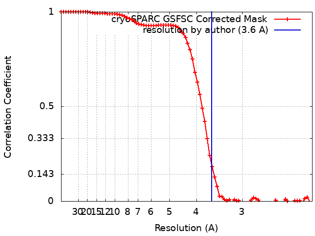
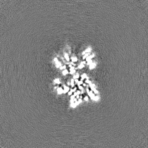
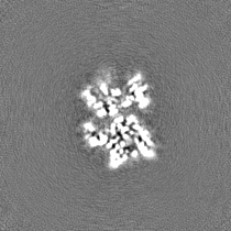
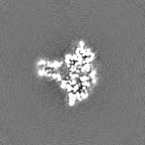
Journal: Proc Natl Acad Sci U S A / Year: 2024 Title: Dual function of LapB (YciM) in regulating lipopolysaccharide synthesis. Authors: Sheng Shu / Yuko Tsutsui / Rajkanwar Nathawat / Wei Mi / Abstract: Levels of lipopolysaccharide (LPS), an essential glycolipid on the surface of most gram-negative bacteria, are tightly controlled-making LPS synthesis a promising target for developing new ...Levels of lipopolysaccharide (LPS), an essential glycolipid on the surface of most gram-negative bacteria, are tightly controlled-making LPS synthesis a promising target for developing new antibiotics. adaptor protein LapB (YciM) plays an important role in regulating LPS synthesis by promoting degradation of LpxC, a deacetylase that catalyzes the first committed step in LPS synthesis. Under conditions where LPS is abundant, LapB recruits LpxC to the AAA+ protease FtsH for degradation. LapB achieves this by simultaneously interacting with FtsH through its transmembrane helix and LpxC through its cytoplasmic domain. Here, we describe a cryo-EM structure of the complex formed between LpxC and the cytoplasmic domain of LapB (LapB). The structure reveals how LapB exploits both its tetratricopeptide repeat (TPR) motifs and rubredoxin domain to interact with LpxC. Through both in vitro and in vivo analysis, we show that mutations at the LapB/LpxC interface prevent LpxC degradation. Unexpectedly, binding to LapB also inhibits the enzymatic activity of LpxC through allosteric effects reminiscent of LpxC activation by MurA in Our findings argue that LapB regulates LPS synthesis in two steps: In the first step, LapB inhibits the activity of LpxC, and in the second step, it commits LpxC to degradation by FtsH.
In the structure databanks used in Yorodumi, some data are registered as the other names, "COVID-19 virus" and "2019-nCoV". Here are the details of the virus and the list of structure data.
Jan 31, 2019. EMDB accession codes are about to change! (news from PDBe EMDB page)
EMDB accession codes are about to change! (news from PDBe EMDB page)
The allocation of 4 digits for EMDB accession codes will soon come to an end. Whilst these codes will remain in use, new EMDB accession codes will include an additional digit and will expand incrementally as the available range of codes is exhausted. The current 4-digit format prefixed with “EMD-” (i.e. EMD-XXXX) will advance to a 5-digit format (i.e. EMD-XXXXX), and so on. It is currently estimated that the 4-digit codes will be depleted around Spring 2019, at which point the 5-digit format will come into force.
The EM Navigator/Yorodumi systems omit the EMD- prefix.
Related info.:Q: What is EMD? / ID/Accession-code notation in Yorodumi/EM Navigator
Yorodumi is a browser for structure data from EMDB, PDB, SASBDB, etc.
This page is also the successor to EM Navigator detail page, and also detail information page/front-end page for Omokage search.
The word "yorodu" (or yorozu) is an old Japanese word meaning "ten thousand". "mi" (miru) is to see.
Related info.:EMDB / PDB / SASBDB / Comparison of 3 databanks / Yorodumi Search / Aug 31, 2016. New EM Navigator & Yorodumi / Yorodumi Papers / Jmol/JSmol / Function and homology information / Changes in new EM Navigator and Yorodumi
 Movie
Movie Controller


 Open data
 Basic information
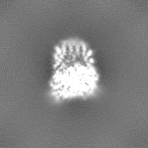
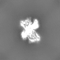
 Map data
 Sample
 Keywords
 Function and homology information
 Authors
 United States, 2 items
  Citation
 Structure visualization
 Downloads & links
 emd_42897.png
 http://ftp.pdbj.org/pub/emdb/structures/EMD-42897
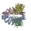


 Z (Sec.)
 Y (Row.)
 X (Col.)


 Sample components

 Processing
 Electron microscopy
 FIELD EMISSION GUN