 Movie
Movie Controller


+ Open data

- Basic information
| Entry | Database: PDB / ID: 8rve | ||||||
|---|---|---|---|---|---|---|---|
| Title | Vimentin intermediate filament | ||||||
 Components Components | Vimentin | ||||||
 Keywords Keywords | STRUCTURAL PROTEIN / vimentin / intermediate filament / cytoskeleton | ||||||
| Function / homology |  Function and homology information Function and homology informationkeratin filament binding / lens fiber cell development / intermediate filament organization / cellular response to muramyl dipeptide / structural constituent of eye lens / astrocyte development / Striated Muscle Contraction / RHOBTB1 GTPase cycle / intermediate filament / microtubule organizing center ...keratin filament binding / lens fiber cell development / intermediate filament organization / cellular response to muramyl dipeptide / structural constituent of eye lens / astrocyte development / Striated Muscle Contraction / RHOBTB1 GTPase cycle / intermediate filament / microtubule organizing center / intermediate filament cytoskeleton / cell leading edge / Bergmann glial cell differentiation / positive regulation of collagen biosynthetic process / Caspase-mediated cleavage of cytoskeletal proteins / phagocytic vesicle / regulation of mRNA stability / Late endosomal microautophagy / structural constituent of cytoskeleton / nuclear matrix / Aggrephagy / cellular response to type II interferon / Chaperone Mediated Autophagy / peroxisome / neuron projection development / double-stranded RNA binding / negative regulation of neuron projection development / scaffold protein binding / Interleukin-4 and Interleukin-13 signaling / cellular response to lipopolysaccharide / molecular adaptor activity / cytoskeleton / axon / protein domain specific binding / focal adhesion / positive regulation of gene expression / extracellular exosome / identical protein binding / plasma membrane / cytosol / cytoplasmSimilarity search - Function | ||||||
| Biological species |  Homo sapiens (human) Homo sapiens (human) | ||||||
| Method | ELECTRON MICROSCOPY / helical reconstruction / cryo EM / Resolution: 7.2 Å | ||||||
 Authors Authors | Eibauer, M. / Medalia, O. | ||||||
| Funding support |  Switzerland, 1items Switzerland, 1items
| ||||||
 Citation Citation | Journal: Nat Struct Mol Biol / Year: 2024 Title: Vimentin filaments integrate low-complexity domains in a complex helical structure. Authors: Matthias Eibauer / Miriam S Weber / Rafael Kronenberg-Tenga / Charlie T Beales / Rajaa Boujemaa-Paterski / Yagmur Turgay / Suganya Sivagurunathan / Julia Kraxner / Sarah Köster / Robert D ...Authors: Matthias Eibauer / Miriam S Weber / Rafael Kronenberg-Tenga / Charlie T Beales / Rajaa Boujemaa-Paterski / Yagmur Turgay / Suganya Sivagurunathan / Julia Kraxner / Sarah Köster / Robert D Goldman / Ohad Medalia /   Abstract: Intermediate filaments (IFs) are integral components of the cytoskeleton. They provide cells with tissue-specific mechanical properties and are involved in numerous cellular processes. Due to their ...Intermediate filaments (IFs) are integral components of the cytoskeleton. They provide cells with tissue-specific mechanical properties and are involved in numerous cellular processes. Due to their intricate architecture, a 3D structure of IFs has remained elusive. Here we use cryo-focused ion-beam milling, cryo-electron microscopy and tomography to obtain a 3D structure of vimentin IFs (VIFs). VIFs assemble into a modular, intertwined and flexible helical structure of 40 α-helices in cross-section, organized into five protofibrils. Surprisingly, the intrinsically disordered head domains form a fiber in the lumen of VIFs, while the intrinsically disordered tails form lateral connections between the protofibrils. Our findings demonstrate how protein domains of low sequence complexity can complement well-folded protein domains to construct a biopolymer with striking mechanical strength and stretchability. | ||||||
| History |
|
- Structure visualization
| Structure viewer | Molecule: MolmilJmol/JSmol |
|---|
- Downloads & links
-Download
| PDBx/mmCIF format | 8rve.cif.gz | 1.8 MB | Display | PDBx/mmCIF format |
|---|---|---|---|---|
| PDB format | pdb8rve.ent.gz | Display | PDB format | |
| PDBx/mmJSON format | 8rve.json.gz | Tree view | PDBx/mmJSON format | |
| Others |  Other downloads Other downloads |
-Validation report
| Arichive directory | https://data.pdbj.org/pub/pdb/validation_reports/rv/8rveftp://data.pdbj.org/pub/pdb/validation_reports/rv/8rve | HTTPS FTP |
|---|
-Related structure data
| Related structure data | 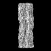 16844MC M: map data used to model this data C: citing same article ( |
|---|---|
| Similar structure data | |
| Other databases |


-Links
 PDBj
PDBj


- Assembly
| Deposited unit | 
|
|---|---|
| 1 |
|
-Components
| #1: Protein | Mass: 53722.582 Da / Num. of mol.: 78 Source method: isolated from a genetically manipulated source Source: (gene. exp.) Homo sapiens (human) / Gene: VIM / Production host:  Escherichia coli (E. coli) / References: UniProt: P08670 Escherichia coli (E. coli) / References: UniProt: P08670 |
|---|
-Experimental details
-Experiment
| Experiment | Method: ELECTRON MICROSCOPY |
|---|---|
| EM experiment | Aggregation state: FILAMENT / 3D reconstruction method: helical reconstruction |
- Sample preparation
| Component | Name: Vimentin intermediate filament / Type: COMPLEX / Entity ID: all / Source: RECOMBINANT |
|---|---|
| Molecular weight | Value: 56 kDa/nm / Experimental value: YES |
| Source (natural) | Organism: Homo sapiens (human) |
| Source (recombinant) | Organism: Escherichia coli (E. coli) |
| Buffer solution | pH: 7.5 |
| Specimen | Embedding applied: NO / Shadowing applied: NO / Staining applied: NO / Vitrification applied: YES |
| Specimen support | Grid material: COPPER / Grid mesh size: 200 divisions/in. / Grid type: Quantifoil R2/1 |
| Vitrification | Instrument: HOMEMADE PLUNGER / Cryogen name: ETHANE |
- Electron microscopy imaging
| Experimental equipment |  Model: Titan Krios / Image courtesy: FEI Company |
|---|---|
| Microscopy | Model: FEI TITAN KRIOS |
| Electron gun | Electron source: FIELD EMISSION GUN / Accelerating voltage: 300 kV / Illumination mode: FLOOD BEAM |
| Electron lens | Mode: BRIGHT FIELDBright-field microscopy / Nominal defocus max: 2800 nm / Nominal defocus min: 800 nm |
| Image recording | Electron dose: 62 e/Å2 / Film or detector model: GATAN K3 BIOQUANTUM (6k x 4k) |
- Processing
| EM software |
| ||||||||||||||||||
|---|---|---|---|---|---|---|---|---|---|---|---|---|---|---|---|---|---|---|---|
| CTF correction | Type: PHASE FLIPPING AND AMPLITUDE CORRECTION | ||||||||||||||||||
| Helical symmerty | Angular rotation/subunit: 73.7308 ° / Axial rise/subunit: 42.461 Å / Axial symmetry: C1 | ||||||||||||||||||
| Particle selection | Num. of particles selected: 1462717 | ||||||||||||||||||
| 3D reconstruction | Resolution: 7.2 Å / Resolution method: FSC 0.143 CUT-OFF / Num. of particles: 236920 / Algorithm: FOURIER SPACE / Symmetry type: HELICAL | ||||||||||||||||||
| Atomic model building | Protocol: FLEXIBLE FIT / Details: PDBDEV_00000212 | ||||||||||||||||||
| Atomic model building | Source name: AlphaFold / Type: in silico model |
