 Movie
Movie Controller


+ Open data

- Basic information
| Entry |  | |||||||||
|---|---|---|---|---|---|---|---|---|---|---|
| Title | Vimentin intermediate filament structure (delta tail) | |||||||||
 Map data Map data | Vimentin intermediate filament structure / delta tail | |||||||||
 Sample Sample |
| |||||||||
 Keywords Keywords |  vimentin / intermediate filament / cytoskeleton / STRUCTURAL PROTEIN vimentin / intermediate filament / cytoskeleton / STRUCTURAL PROTEIN | |||||||||
| Function / homology |  Function and homology information Function and homology informationkeratin filament binding / lens fiber cell development / intermediate filament organization / cellular response to muramyl dipeptide / structural constituent of eye lens / astrocyte development / Striated Muscle Contraction / RHOBTB1 GTPase cycle / intermediate filament / microtubule organizing center ...keratin filament binding / lens fiber cell development / intermediate filament organization / cellular response to muramyl dipeptide / structural constituent of eye lens / astrocyte development / Striated Muscle Contraction / RHOBTB1 GTPase cycle / intermediate filament / microtubule organizing center / intermediate filament cytoskeleton / cell leading edge / Bergmann glial cell differentiation / positive regulation of collagen biosynthetic process / Caspase-mediated cleavage of cytoskeletal proteins / phagocytic vesicle / regulation of mRNA stability / Late endosomal microautophagy / structural constituent of cytoskeleton / nuclear matrix / Aggrephagy / cellular response to type II interferon / Chaperone Mediated Autophagy / peroxisome / neuron projection development / double-stranded RNA binding / negative regulation of neuron projection development / scaffold protein binding / Interleukin-4 and Interleukin-13 signaling / cellular response to lipopolysaccharide / molecular adaptor activity / cytoskeleton / axon / protein domain specific binding / focal adhesion / positive regulation of gene expression / extracellular exosome / identical protein binding / plasma membrane / cytosol / cytoplasmSimilarity search - Function | |||||||||
| Biological species |  Homo sapiens (human) Homo sapiens (human) | |||||||||
| Method | helical reconstruction / cryo EM / Resolution: 14.0 Å | |||||||||
 Authors Authors | Eibauer M / Medalia O | |||||||||
| Funding support |  Switzerland, 1 items Switzerland, 1 items
| |||||||||
 Citation Citation | Journal: Nat Struct Mol Biol / Year: 2024 Title: Vimentin filaments integrate low-complexity domains in a complex helical structure. Authors: Matthias Eibauer / Miriam S Weber / Rafael Kronenberg-Tenga / Charlie T Beales / Rajaa Boujemaa-Paterski / Yagmur Turgay / Suganya Sivagurunathan / Julia Kraxner / Sarah Köster / Robert D ...Authors: Matthias Eibauer / Miriam S Weber / Rafael Kronenberg-Tenga / Charlie T Beales / Rajaa Boujemaa-Paterski / Yagmur Turgay / Suganya Sivagurunathan / Julia Kraxner / Sarah Köster / Robert D Goldman / Ohad Medalia /   Abstract: Intermediate filaments (IFs) are integral components of the cytoskeleton. They provide cells with tissue-specific mechanical properties and are involved in numerous cellular processes. Due to their ...Intermediate filaments (IFs) are integral components of the cytoskeleton. They provide cells with tissue-specific mechanical properties and are involved in numerous cellular processes. Due to their intricate architecture, a 3D structure of IFs has remained elusive. Here we use cryo-focused ion-beam milling, cryo-electron microscopy and tomography to obtain a 3D structure of vimentin IFs (VIFs). VIFs assemble into a modular, intertwined and flexible helical structure of 40 α-helices in cross-section, organized into five protofibrils. Surprisingly, the intrinsically disordered head domains form a fiber in the lumen of VIFs, while the intrinsically disordered tails form lateral connections between the protofibrils. Our findings demonstrate how protein domains of low sequence complexity can complement well-folded protein domains to construct a biopolymer with striking mechanical strength and stretchability. | |||||||||
| History |
|
- Structure visualization
| Supplemental images |
|---|

- Downloads & links
-EMDB archive
| Map data | emd_19563.map.gz | 196.6 MB | EMDB map data format | |
|---|---|---|---|---|
| Header (meta data) | emd-19563-v30.xmlemd-19563.xml | 15 KB 15 KB | Display Display | EMDB header |
| FSC (resolution estimation) | emd_19563_fsc.xml | 13.6 KB | Display | FSC data file |
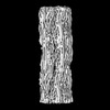
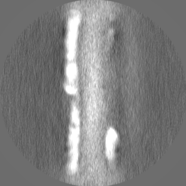
| Images | 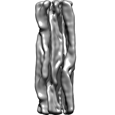 emd_19563.png emd_19563.png | 55 KB | ||
| Masks | emd_19563_msk_1.map | 209.3 MB | Mask map | |
| Filedesc metadata | emd-19563.cif.gz | 5.1 KB | ||
| Others | emd_19563_half_map_1.map.gzemd_19563_half_map_2.map.gz | 164.9 MB 164.8 MB | ||
| Archive directory |  http://ftp.pdbj.org/pub/emdb/structures/EMD-19563ftp://ftp.pdbj.org/pub/emdb/structures/EMD-19563 http://ftp.pdbj.org/pub/emdb/structures/EMD-19563ftp://ftp.pdbj.org/pub/emdb/structures/EMD-19563 | HTTPS FTP |
-Related structure data


-Links
| EMDB pages | EMDB (EBI/PDBe) / EMDataResource |
|---|---|
| Related items in Molecule of the Month |


-Map
| File | Download / File: emd_19563.map.gz / Format: CCP4 / Size: 209.3 MB / Type: IMAGE STORED AS FLOATING POINT NUMBER (4 BYTES) | ||||||||||||||||||||
|---|---|---|---|---|---|---|---|---|---|---|---|---|---|---|---|---|---|---|---|---|---|
| Annotation | Vimentin intermediate filament structure / delta tail | ||||||||||||||||||||
| Voxel size | X=Y=Z: 1.0021 Å | ||||||||||||||||||||
| Density |
| ||||||||||||||||||||
| Symmetry | Space group: 1 | ||||||||||||||||||||
| Details | EMDB XML:
|
-Supplemental data
-Mask #1
| File | emd_19563_msk_1.map | ||||||||||||
|---|---|---|---|---|---|---|---|---|---|---|---|---|---|
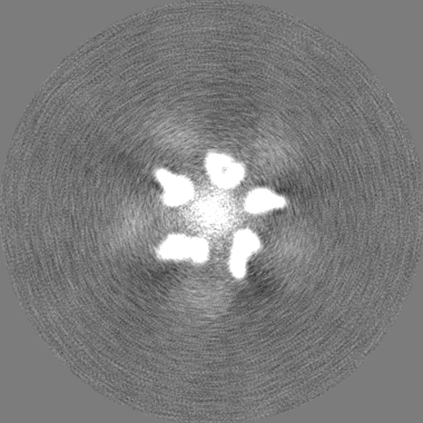
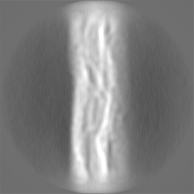
| Projections & Slices |
| ||||||||||||
| Density Histograms |
 Z
Z Y
Y X
X


-Half map: Vimentin intermediate filament structure / delta tail / half map 1
| File | emd_19563_half_map_1.map | ||||||||||||
|---|---|---|---|---|---|---|---|---|---|---|---|---|---|
| Annotation | Vimentin intermediate filament structure / delta tail / half map 1 | ||||||||||||
| Projections & Slices |
| ||||||||||||
| Density Histograms |


-Half map: Vimentin intermediate filament structure / delta tail / half map 2
| File | emd_19563_half_map_2.map | ||||||||||||
|---|---|---|---|---|---|---|---|---|---|---|---|---|---|
| Annotation | Vimentin intermediate filament structure / delta tail / half map 2 | ||||||||||||
| Projections & Slices |
| ||||||||||||
| Density Histograms |


- Sample components
-Entire : Vimentin intermediate filament (delta tail)
| Entire | Name: Vimentin intermediate filament (delta tail) |
|---|---|
| Components |
|
-Supramolecule #1: Vimentin intermediate filament (delta tail)
| Supramolecule | Name: Vimentin intermediate filament (delta tail) / type: complex / ID: 1 / Parent: 0 / Macromolecule list: all |
|---|---|
| Source (natural) | Organism: Homo sapiens (human) |
-Macromolecule #1: Vimentin (delta tail)
| Macromolecule | Name: Vimentin (delta tail) / type: protein_or_peptide / ID: 1 / Enantiomer: LEVO |
|---|---|
| Source (natural) | Organism: Homo sapiens (human) |
| Recombinant expression | Organism:  Escherichia coli (E. coli) Escherichia coli (E. coli) |
| Sequence | String: MSTRSVSSSS YRRMFGGPGT ASRPSSSRSY VTTSTRTYSL GSALRPSTSR SLYASSPGGV YATRSSAVRL RSSVPGVRLL QDSVDFSLAD AINTEFKNTR TNEKVELQEL NDRFANYIDK VRFLEQQNKI LLAELEQLKG QGKSRLGDLY EEEMRELRRQ VDQLTNDKAR ...String: MSTRSVSSSS YRRMFGGPGT ASRPSSSRSY VTTSTRTYSL GSALRPSTSR SLYASSPGGV YATRSSAVRL RSSVPGVRLL QDSVDFSLAD AINTEFKNTR TNEKVELQEL NDRFANYIDK VRFLEQQNKI LLAELEQLKG QGKSRLGDLY EEEMRELRRQ VDQLTNDKAR VEVERDNLAE DIMRLREKLQ EEMLQREEAE NTLQSFRQDV DNASLARLDL ERKVESLQEE IAFLKKLHEE EIQELQAQIQ EQHVQIDVDV SKPDLTAALR DVRQQYESVA AKNLQEAEEW YKSKFADLSE AANRNNDALR QAKQESTEYR RQVQSLTCEV DALKGTNESL ERQMREMEEN FAVEAANYQD TIGRLQDEIQ NMKEEMARHL REYQDLLNVK MALDIEIATY RKLLEGEES UniProtKB: Vimentin |
-Experimental details
-Structure determination
| Method | cryo EM |
|---|---|
 Processing Processing | helical reconstruction |
| Aggregation state | filament |
-Sample preparation
| Buffer | pH: 7.5 |
|---|---|
| Grid | Model: Quantifoil R2/1 / Material: COPPER / Mesh: 200 / Support film - Material: CARBON / Support film - topology: HOLEY |
| Vitrification | Cryogen name: ETHANE / Instrument: HOMEMADE PLUNGER |
- Electron microscopy
| Microscope | FEI TITAN KRIOS |
|---|---|
| Electron beam | Acceleration voltage: 300 kV / Electron source: FIELD EMISSION GUN |
| Electron optics | Illumination mode: FLOOD BEAM / Imaging mode: BRIGHT FIELDBright-field microscopy / Nominal defocus max: 2.8000000000000003 µm / Nominal defocus min: 0.8 µm |
| Image recording | Film or detector model: GATAN K3 BIOQUANTUM (6k x 4k) / Average electron dose: 62.0 e/Å2 |
| Experimental equipment |  Model: Titan Krios / Image courtesy: FEI Company |
-Image processing
| Segment selection | Number selected: 1019393 / Software - Name: crYOLO |
|---|---|
| Startup model | Type of model: OTHER |
| Final angle assignment | Type: NOT APPLICABLE |
| Final reconstruction | Applied symmetry - Helical parameters - Δz: 42.5 Å Applied symmetry - Helical parameters - Δ&Phi: 73.7 ° Applied symmetry - Helical parameters - Axial symmetry: C1 (asymmetric) Algorithm: FOURIER SPACE / Resolution.type: BY AUTHOR / Resolution: 14.0 Å / Resolution method: FSC 0.143 CUT-OFF / Software - Name: RELION / Number images used: 143270 |
| FSC plot (resolution estimation) | 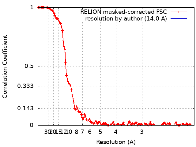 |
