 Movie
Movie Controller

[English] 日本語
 Yorodumi
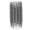
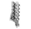
- PDB-8qv2: Structure of the native y-Tubulin Ring Complex (yTuRC) capping mi... -
+ Open data

- Basic information
| Entry | Database: PDB / ID: 8qv2 | ||||||||||||
|---|---|---|---|---|---|---|---|---|---|---|---|---|---|
| Title | Structure of the native y-Tubulin Ring Complex (yTuRC) capping microtubule minus ends at the spindle pole body | ||||||||||||
 Components Components |
| ||||||||||||
 Keywords Keywords |  CELL CYCLE / Microtubule nucleation / MTOC / y-tubulin / SPB CELL CYCLE / Microtubule nucleation / MTOC / y-tubulin / SPB | ||||||||||||
| Function / homology |  Function and homology informationCilium Assembly / nuclear migration by microtubule mediated pushing forces / nuclear division / Sealing of the nuclear envelope (NE) by ESCRT-III / nuclear migration along microtubule / homologous chromosome segregation / gamma-tubulin complex / Platelet degranulation / microtubule nucleation / spindle pole body ...Cilium Assembly / nuclear migration by microtubule mediated pushing forces / nuclear division / Sealing of the nuclear envelope (NE) by ESCRT-III / nuclear migration along microtubule / homologous chromosome segregation / gamma-tubulin complex / Platelet degranulation / microtubule nucleation / spindle pole body / gamma-tubulin binding / tubulin complex / mitotic sister chromatid segregation / microtubule-based process / cytoplasmic microtubule organization / nuclear periphery / Hydrolases; Acting on acid anhydrides; Acting on GTP to facilitate cellular and subcellular movement / structural constituent of cytoskeleton / spindle / microtubule cytoskeleton organization / spindle pole / mitotic cell cycle / microtubule / hydrolase activity / GTPase activity / GTP binding / metal ion binding / nucleus / cytoplasm Function and homology informationCilium Assembly / nuclear migration by microtubule mediated pushing forces / nuclear division / Sealing of the nuclear envelope (NE) by ESCRT-III / nuclear migration along microtubule / homologous chromosome segregation / gamma-tubulin complex / Platelet degranulation / microtubule nucleation / spindle pole body ...Cilium Assembly / nuclear migration by microtubule mediated pushing forces / nuclear division / Sealing of the nuclear envelope (NE) by ESCRT-III / nuclear migration along microtubule / homologous chromosome segregation / gamma-tubulin complex / Platelet degranulation / microtubule nucleation / spindle pole body / gamma-tubulin binding / tubulin complex / mitotic sister chromatid segregation / microtubule-based process / cytoplasmic microtubule organization / nuclear periphery / Hydrolases; Acting on acid anhydrides; Acting on GTP to facilitate cellular and subcellular movement / structural constituent of cytoskeleton / spindle / microtubule cytoskeleton organization / spindle pole / mitotic cell cycle / microtubule / hydrolase activity / GTPase activity / GTP binding / metal ion binding / nucleus / cytoplasmSimilarity search - Function | ||||||||||||
| Biological species |  Saccharomyces cerevisiae (brewer's yeast) Saccharomyces cerevisiae (brewer's yeast) | ||||||||||||
| Method | ELECTRON MICROSCOPY / subtomogram averaging / cryo EM / Resolution: 9.2 Å | ||||||||||||
 Authors Authors | Dendooven, T. / Yatskevich, S. / Burt, A. / Bellini, D. / Kilmartin, J. / Barford, D. | ||||||||||||
| Funding support |  United Kingdom, United Kingdom,  Germany, 3items Germany, 3items
| ||||||||||||
 Citation Citation | Journal: Nat Struct Mol Biol / Year: 2024 Title: Structure of the native γ-tubulin ring complex capping spindle microtubules. Authors: Tom Dendooven / Stanislau Yatskevich / Alister Burt / Zhuo A Chen / Dom Bellini / Juri Rappsilber / John V Kilmartin / David Barford /  Abstract: Microtubule (MT) filaments, composed of α/β-tubulin dimers, are fundamental to cellular architecture, function and organismal development. They are nucleated from MT organizing centers by the ...Microtubule (MT) filaments, composed of α/β-tubulin dimers, are fundamental to cellular architecture, function and organismal development. They are nucleated from MT organizing centers by the evolutionarily conserved γ-tubulin ring complex (γTuRC). However, the molecular mechanism of nucleation remains elusive. Here we used cryo-electron tomography to determine the structure of the native γTuRC capping the minus end of a MT in the context of enriched budding yeast spindles. In our structure, γTuRC presents a ring of γ-tubulin subunits to seed nucleation of exclusively 13-protofilament MTs, adopting an active closed conformation to function as a perfect geometric template for MT nucleation. Our cryo-electron tomography reconstruction revealed that a coiled-coil protein staples the first row of α/β-tubulin of the MT to alternating positions along the γ-tubulin ring of γTuRC. This positioning of α/β-tubulin onto γTuRC suggests a role for the coiled-coil protein in augmenting γTuRC-mediated MT nucleation. Based on our results, we describe a molecular model for budding yeast γTuRC activation and MT nucleation. | ||||||||||||
| History |
|
- Structure visualization
| Structure viewer | Molecule: MolmilJmol/JSmol |
|---|
- Downloads & links
-Download
| PDBx/mmCIF format | 8qv2.cif.gz | 5.5 MB | Display | PDBx/mmCIF format |
|---|---|---|---|---|
| PDB format | pdb8qv2.ent.gz | Display | PDB format | |
| PDBx/mmJSON format | 8qv2.json.gz | Tree view | PDBx/mmJSON format | |
| Others |  Other downloads Other downloads |
-Validation report
| Arichive directory | https://data.pdbj.org/pub/pdb/validation_reports/qv/8qv2ftp://data.pdbj.org/pub/pdb/validation_reports/qv/8qv2 | HTTPS FTP |
|---|
-Related structure data
| Related structure data | 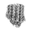 18665MC 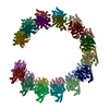 8qv0C 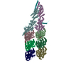 8qv3C M: map data used to model this data C: citing same article ( |
|---|---|
| Similar structure data |
-Links
 PDBj
PDBj


- Assembly
| Deposited unit | 
|
|---|---|
| 1 |
|
-Components
-Protein , 4 types, 62 molecules bcdefghijklmnaAdAcAfAeAhAgAjAiAlAkAnAmAbApAoAr...
| #1: Protein | Mass: 52671.188 Da / Num. of mol.: 14 / Source method: isolated from a natural source / Source: (natural) Saccharomyces cerevisiae (brewer's yeast) / References: UniProt: A0A8H4BZN3#4: Protein | Mass: 49853.867 Da / Num. of mol.: 17 / Source method: isolated from a natural source / Source: (natural) Saccharomyces cerevisiae (brewer's yeast) / References: UniProt: P09733#5: Protein | Mass: 50967.457 Da / Num. of mol.: 17 / Source method: isolated from a natural source / Source: (natural) Saccharomyces cerevisiae (brewer's yeast) / References: UniProt: A0A6A5PXT5#7: Protein | Mass: 5720.042 Da / Num. of mol.: 14 / Source method: isolated from a natural source / Source: (natural) Saccharomyces cerevisiae (brewer's yeast) |
|---|
-Spindle pole body ... , 3 types, 28 molecules CEGIKMODFHJLNPScSdSeSfSgShSiSjSkSlSaSbSmSn
| #2: Protein | Mass: 96940.594 Da / Num. of mol.: 7 / Source method: isolated from a natural source / Source: (natural) Saccharomyces cerevisiae (brewer's yeast) / References: UniProt: A0A8H4C290#3: Protein | Mass: 98336.211 Da / Num. of mol.: 7 / Source method: isolated from a natural source / Source: (natural) Saccharomyces cerevisiae (brewer's yeast) / References: UniProt: A0A8H4BVY6#6: Protein | Mass: 111987.125 Da / Num. of mol.: 14 / Source method: isolated from a natural source / Source: (natural) Saccharomyces cerevisiae (brewer's yeast) / References: UniProt: A0A8H8UNQ3 |
|---|
-Non-polymers , 3 types, 21 molecules 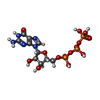
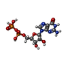

| #8: Chemical | ChemComp-GTP / Guanosine triphosphate Mass: 523.180 Da / Num. of mol.: 13 / Source method: obtained synthetically / Formula: C10H16N5O14P3 / Comment: GTP, energy-carrying molecule*YM Mass: 523.180 Da / Num. of mol.: 13 / Source method: obtained synthetically / Formula: C10H16N5O14P3 / Comment: GTP, energy-carrying molecule*YM#9: Chemical | ChemComp-GDP / | Guanosine diphosphate Type: RNA linking / Mass: 443.201 Da / Num. of mol.: 1 / Source method: obtained synthetically / Formula: C10H15N5O11P2 / Comment: GDP, energy-carrying molecule*YM Type: RNA linking / Mass: 443.201 Da / Num. of mol.: 1 / Source method: obtained synthetically / Formula: C10H15N5O11P2 / Comment: GDP, energy-carrying molecule*YM#10: Water | ChemComp-HOH / | WaterMass: 18.015 Da / Num. of mol.: 7 / Source method: isolated from a natural source / Formula: H2O |
|---|
-Details
| Has ligand of interest | N |
|---|
-Experimental details
-Experiment
| Experiment | Method: ELECTRON MICROSCOPY |
|---|---|
| EM experiment | Aggregation state: PARTICLE / 3D reconstruction method: subtomogram averaging |
- Sample preparation
| Component | Name: y-Tubulin Ring Complex capping the microtubule minus end Type: ORGANELLE OR CELLULAR COMPONENT / Entity ID: #1-#7 / Source: NATURAL |
|---|---|
| Molecular weight | Experimental value: NO |
| Source (natural) | Organism: Saccharomyces cerevisiae (brewer's yeast) |
| Buffer solution | pH: 6.53 |
| Specimen | Embedding applied: NO / Shadowing applied: NO / Staining applied: NO / Vitrification applied: YES |
| Vitrification | Cryogen name: ETHANE |
- Electron microscopy imaging
| Experimental equipment |  Model: Titan Krios / Image courtesy: FEI Company |
|---|---|
| Microscopy | Model: FEI TITAN KRIOS |
| Electron gun | Electron source: FIELD EMISSION GUN / Accelerating voltage: 300 kV / Illumination mode: SPOT SCAN |
| Electron lens | Mode: BRIGHT FIELDBright-field microscopy / Nominal defocus max: 4500 nm / Nominal defocus min: 2000 nm / C2 aperture diameter: 50 µm |
| Image recording | Electron dose: 3 e/Å2 / Avg electron dose per subtomogram: 123 e/Å2 / Film or detector model: GATAN K3 (6k x 4k) |
- Processing
| EM software | Name: PHENIX / Version: 1.17.1_3660: / Category: model refinement | ||||||||||||||||||||||||
|---|---|---|---|---|---|---|---|---|---|---|---|---|---|---|---|---|---|---|---|---|---|---|---|---|---|
| CTF correction | Type: PHASE FLIPPING AND AMPLITUDE CORRECTION | ||||||||||||||||||||||||
| Symmetry | Point symmetry: C1 (asymmetric) | ||||||||||||||||||||||||
| 3D reconstruction | Resolution: 9.2 Å / Resolution method: FSC 0.143 CUT-OFF / Num. of particles: 7910 / Symmetry type: POINT | ||||||||||||||||||||||||
| EM volume selection | Num. of tomograms: 364 / Num. of volumes extracted: 31720 | ||||||||||||||||||||||||
| Refine LS restraints |
|
