ムービー
ムービー コントローラー


+ データを開く

- 基本情報
基本情報
| 登録情報 | データベース: EMDB / ID: EMD-1531 | |||||||||
|---|---|---|---|---|---|---|---|---|---|---|
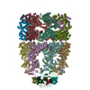
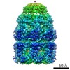
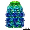
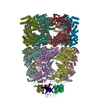
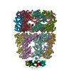
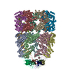
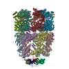
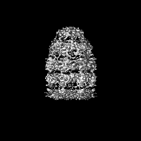
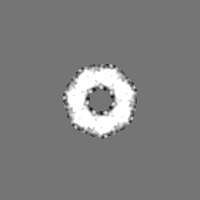
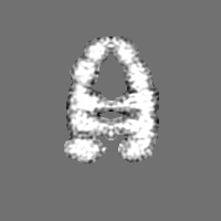
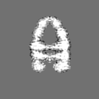
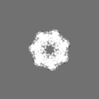
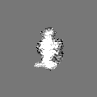
| タイトル | Three-dimensional structure of Aquifex aeolicus co-chaperonin protein 10 complexed with GroEL and ADP by cryo-EM | |||||||||
 マップデータ マップデータ | none | |||||||||
 試料 試料 |
| |||||||||
 キーワード キーワード | co-chaperonin / hyper-thermophile / conformational heterogeneity /  electron cryomicroscopy (低温電子顕微鏡法) electron cryomicroscopy (低温電子顕微鏡法) | |||||||||
| 機能・相同性 | :  機能・相同性情報 機能・相同性情報 | |||||||||
| 生物種 |  Escherichia coli (大腸菌) / Escherichia coli (大腸菌) /  Aquifex aeolicus (バクテリア) Aquifex aeolicus (バクテリア) | |||||||||
| 手法 | 単粒子再構成法 / クライオ電子顕微鏡法 / ネガティブ染色法 / 解像度: 8.0 Å | |||||||||
 データ登録者 データ登録者 | Chen DH / Luke K / Zhang J / Chiu W / Wittung-Stafshede P | |||||||||
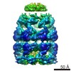
 引用 引用 | ジャーナル: J Mol Biol / 年: 2008 タイトル: Location and flexibility of the unique C-terminal tail of Aquifex aeolicus co-chaperonin protein 10 as derived by cryo-electron microscopy and biophysical techniques. 著者: Dong-Hua Chen / Kathryn Luke / Junjie Zhang / Wah Chiu / Pernilla Wittung-Stafshede /  要旨: Co-chaperonin protein 10 (cpn10, GroES in Escherichia coli) is a ring-shaped heptameric protein that facilitates substrate folding when in complex with cpn60 (GroEL in E. coli). The cpn10 from the ...Co-chaperonin protein 10 (cpn10, GroES in Escherichia coli) is a ring-shaped heptameric protein that facilitates substrate folding when in complex with cpn60 (GroEL in E. coli). The cpn10 from the hyperthermophilic, ancient bacterium Aquifex aeolicus (Aacpn10) has a 25-residue C-terminal extension in each monomer not found in any other cpn10 protein. Earlier in vitro work has shown that this tail is not needed for heptamer assembly or protein function. Without the tail, however, the heptamers (Aacpn10del-25) readily aggregate into fibrillar stacked rings. To explain this phenomenon, we performed binding experiments with a peptide construct of the tail to establish its specificity for Aacpn10del-25 and used cryo-electron microscopy to determine the three-dimensional (3D) structure of the GroEL-Aacpn10-ADP complex at an 8-A resolution. We found that the GroEL-Aacpn10 structure is similar to the GroEL-GroES structure at this resolution, suggesting that Aacpn10 has molecular interactions with cpn60 similar to other cpn10s. The cryo-electron microscopy density map does not directly reveal the density of the Aacpn10 25-residue tail. However, the 3D statistical variance coefficient map computed from multiple 3D reconstructions with randomly selected particle images suggests that the tail is located at the Aacpn10 monomer-monomer interface and extends toward the cis-ring apical domain of GroEL. The tail at this location does not block the formation of a functional co-chaperonin/chaperonin complex but limits self-aggregation into linear fibrils at high temperatures. In addition, the 3D variance coefficient map identifies several regions inside the GroEL-Aacpn10 complex that have flexible conformations. This observation is in full agreement with the structural properties of an effective chaperonin. | |||||||||
| 履歴 |
|
- 構造の表示
構造の表示
| ムービー |
ムービービューア |
|---|---|
| 構造ビューア | EMマップ: SurfViewMolmilJmol/JSmol |
| 添付画像 |

- ダウンロードとリンク
-EMDBアーカイブ
| マップデータ | emd_1531.map.gz | 2.6 MB | EMDBマップデータ形式 | |
|---|---|---|---|---|
| ヘッダ (付随情報) | emd-1531-v30.xmlemd-1531.xml | 12.5 KB 12.5 KB | 表示 表示 | EMDBヘッダ |
| 画像 |  1531.gif 1531.gif | 218.5 KB | ||
| アーカイブディレクトリ |  http://ftp.pdbj.org/pub/emdb/structures/EMD-1531ftp://ftp.pdbj.org/pub/emdb/structures/EMD-1531 http://ftp.pdbj.org/pub/emdb/structures/EMD-1531ftp://ftp.pdbj.org/pub/emdb/structures/EMD-1531 | HTTPS FTP |
-関連構造データ
-リンク
| EMDBのページ | EMDB (EBI/PDBe) / EMDataResource |
|---|
-マップ
| ファイル | ダウンロード / ファイル: emd_1531.map.gz / 形式: CCP4 / 大きさ: 29.8 MB / タイプ: IMAGE STORED AS FLOATING POINT NUMBER (4 BYTES) | ||||||||||||||||||||||||||||||||||||||||||||||||||||||||||||||||||||
|---|---|---|---|---|---|---|---|---|---|---|---|---|---|---|---|---|---|---|---|---|---|---|---|---|---|---|---|---|---|---|---|---|---|---|---|---|---|---|---|---|---|---|---|---|---|---|---|---|---|---|---|---|---|---|---|---|---|---|---|---|---|---|---|---|---|---|---|---|---|
| 注釈 | none | ||||||||||||||||||||||||||||||||||||||||||||||||||||||||||||||||||||
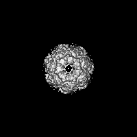
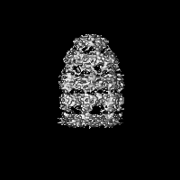
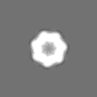
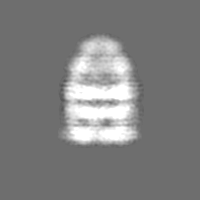
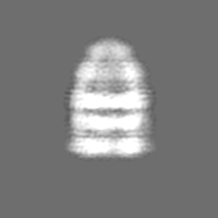
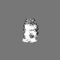
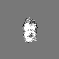
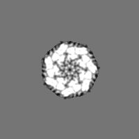
| 投影像・断面図 | 画像のコントロール
画像は Spider により作成 | ||||||||||||||||||||||||||||||||||||||||||||||||||||||||||||||||||||
| ボクセルのサイズ | X=Y=Z: 1.795 Å | ||||||||||||||||||||||||||||||||||||||||||||||||||||||||||||||||||||
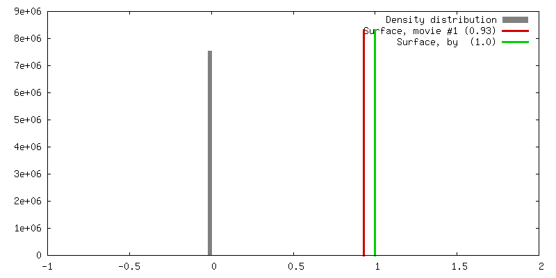
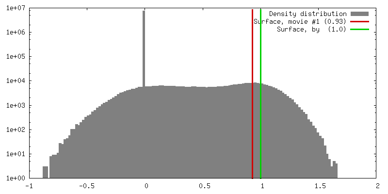
| 密度 |
| ||||||||||||||||||||||||||||||||||||||||||||||||||||||||||||||||||||
| 対称性 | 空間群: 1 | ||||||||||||||||||||||||||||||||||||||||||||||||||||||||||||||||||||
| 詳細 | EMDB XML:
CCP4マップ ヘッダ情報:
| ||||||||||||||||||||||||||||||||||||||||||||||||||||||||||||||||||||
 Z (Sec.)
 Y (Row.)
 X (Col.)


-添付データ
- 試料の構成要素
-全体 : Aacpn10 capped to one end of GroEL under Mg-ADP
| 全体 | 名称: Aacpn10 capped to one end of GroEL under Mg-ADP |
|---|---|
| 要素 |
|
-超分子 #1000: Aacpn10 capped to one end of GroEL under Mg-ADP
| 超分子 | 名称: Aacpn10 capped to one end of GroEL under Mg-ADP / タイプ: sample / ID: 1000 詳細: Aacpn10 was incubated with GroEL in 50 mM Tris-HCl, 30 mM MgCl2, 2 mM ADP, pH 7.5 at 37 Celsius for 1 hour 集合状態: One heptamer of Aacpn10 and seven ADP bind to one heptamer of GroEL Number unique components: 2 |
|---|---|
| 分子量 | 実験値: 900 KDa / 理論値: 900 KDa |
-分子 #1: Chaperonin
| 分子 | 名称: Chaperonin / タイプ: protein_or_peptide / ID: 1 / Name.synonym: GroEL / コピー数: 1 / 集合状態: heptamer / 組換発現: Yes |
|---|---|
| 由来(天然) | 生物種: Escherichia coli (大腸菌) |
| 分子量 | 実験値: 800 KDa / 理論値: 800 KDa |
| 組換発現 | 生物種: Escherichia coli (大腸菌) / 組換プラスミド: pMESL |
| 配列 | InterPro: INTERPRO: IPR012723 |
-分子 #2: Aquifex aeolicus co-chaperonin 10
| 分子 | 名称: Aquifex aeolicus co-chaperonin 10 / タイプ: protein_or_peptide / ID: 2 / Name.synonym: Aacpn10 / コピー数: 1 / 集合状態: heptamer / 組換発現: Yes |
|---|---|
| 由来(天然) | 生物種: Aquifex aeolicus (バクテリア) / 株: AQ2199 / 組織: Bacterial / 細胞: Aquifex aeolicus / 細胞中の位置: Cytosol |
| 分子量 | 実験値: 100 KDa / 理論値: 100 KDa |
| 組換発現 | 生物種: Escherichia coli (大腸菌) / 組換プラスミド: pET24c |
-実験情報
-構造解析
| 手法 | ネガティブ染色法, クライオ電子顕微鏡法 |
|---|---|
 解析 解析 | 単粒子再構成法 |
| 試料の集合状態 | particle |
-試料調製
| 濃度 | 4 mg/mL |
|---|---|
| 緩衝液 | pH: 7.5 / 詳細: 50 mM Tris-HCl, 30 mM MgCl2 |
| 染色 | タイプ: NEGATIVE 詳細: The sample solution was blotted for 2.5 s before submersion in liquid ethane cooled by liquid nitrogen using FEI Vitrobot |
| グリッド | 詳細: 400 mesh copper grid (R1.2/1.3 Quantifoil) |
| 凍結 | 凍結剤: ETHANE / チャンバー内湿度: 95 % / チャンバー内温度: 4.2 K / 装置: OTHER / 詳細: Vitrification instrument: Vitrobot / 手法: blot for 2.5 s before plunging |
- 電子顕微鏡法
| 顕微鏡 | JEOL 3000SFF |
|---|---|
| 電子線 | 加速電圧: 300 kV / 電子線源: FIELD EMISSION GUN |
| 電子光学系 | 照射モード: FLOOD BEAM / 撮影モード: BRIGHT FIELDBright-field microscopy / Cs: 1.6 mm / 最大 デフォーカス(公称値): 3.4 µm / 最小 デフォーカス(公称値): 0.86 µm / 倍率(公称値): 50000 |
| 試料ステージ | 試料ホルダー: top-entry / 試料ホルダーモデル: OTHER |
| 温度 | 最低: 4.2 K / 最高: 4.2 K / 平均: 4.2 K |
| アライメント法 | Legacy - 非点収差: objective lens astigmatism was corrected at 400,000 times magnification |
| 詳細 | Yoshi MDS box for low-dose imaging |
| 日付 | 2007年2月16日 |
| 撮影 | カテゴリ: FILM / フィルム・検出器のモデル: KODAK SO-163 FILM デジタル化 - スキャナー: NIKON SUPER COOLSCAN 9000 デジタル化 - サンプリング間隔: 6.35 µm / 実像数: 720 / 平均電子線量: 36 e/Å2 詳細: The particle images were averaged by 2 to give 1.91 Angstrom per pixel. The final corrected pixel size for the 3D density map is 1.8 Angstrom per pixel. ビット/ピクセル: 8 |
-画像解析
| CTF補正 | 詳細: each micrograph |
|---|---|
| 最終 2次元分類 | クラス数: 514 |
| 最終 角度割当 | 詳細: EMAN |
| 最終 再構成 | 想定した対称性 - 点群: C7 (7回回転対称) / アルゴリズム: OTHER / 解像度のタイプ: BY AUTHOR / 解像度: 8.0 Å / 解像度の算出法: FSC 0.5 CUT-OFF / ソフトウェア - 名称: EMAN 詳細: The final map was calculated from the class averages and was filtered to show the subnanometer resolution features. 使用した粒子像数: 10772 |
| 詳細 | The ratio of GroEL monomers to Aacpn10 monomers was about 1 |
-原子モデル構築 1
| 初期モデル | PDB ID: Chain - Chain ID: A |
|---|---|
| ソフトウェア | 名称: UROX |
| 詳細 | PDBEntryID_givenInChain. Protocol: domain-based rigid body. UROX was used to perform the domain-as-rigid-body fitting of the GroEL-GroES-ADP crystal structure into the cryo-EM density map of the GroEL-Aacpn10-ADP complex |
| 精密化 | 空間: REAL / プロトコル: RIGID BODY FIT / 当てはまり具合の基準: cross correlation |