 Movie
Movie Controller
 Structure viewers
 About Yorodumi Papers


+Search query
-Structure paper
| Title | Location and flexibility of the unique C-terminal tail of Aquifex aeolicus co-chaperonin protein 10 as derived by cryo-electron microscopy and biophysical techniques. |
|---|---|
| Journal, issue, pages | J Mol Biol, Vol. 381, Issue 3, Page 707-717, Year 2008 |
| Publish date | Sep 5, 2008 |
 Authors Authors | Dong-Hua Chen / Kathryn Luke / Junjie Zhang / Wah Chiu / Pernilla Wittung-Stafshede /  |
| PubMed Abstract | Co-chaperonin protein 10 (cpn10, GroES in Escherichia coli) is a ring-shaped heptameric protein that facilitates substrate folding when in complex with cpn60 (GroEL in E. coli). The cpn10 from the ...Co-chaperonin protein 10 (cpn10, GroES in Escherichia coli) is a ring-shaped heptameric protein that facilitates substrate folding when in complex with cpn60 (GroEL in E. coli). The cpn10 from the hyperthermophilic, ancient bacterium Aquifex aeolicus (Aacpn10) has a 25-residue C-terminal extension in each monomer not found in any other cpn10 protein. Earlier in vitro work has shown that this tail is not needed for heptamer assembly or protein function. Without the tail, however, the heptamers (Aacpn10del-25) readily aggregate into fibrillar stacked rings. To explain this phenomenon, we performed binding experiments with a peptide construct of the tail to establish its specificity for Aacpn10del-25 and used cryo-electron microscopy to determine the three-dimensional (3D) structure of the GroEL-Aacpn10-ADP complex at an 8-A resolution. We found that the GroEL-Aacpn10 structure is similar to the GroEL-GroES structure at this resolution, suggesting that Aacpn10 has molecular interactions with cpn60 similar to other cpn10s. The cryo-electron microscopy density map does not directly reveal the density of the Aacpn10 25-residue tail. However, the 3D statistical variance coefficient map computed from multiple 3D reconstructions with randomly selected particle images suggests that the tail is located at the Aacpn10 monomer-monomer interface and extends toward the cis-ring apical domain of GroEL. The tail at this location does not block the formation of a functional co-chaperonin/chaperonin complex but limits self-aggregation into linear fibrils at high temperatures. In addition, the 3D variance coefficient map identifies several regions inside the GroEL-Aacpn10 complex that have flexible conformations. This observation is in full agreement with the structural properties of an effective chaperonin. |
 External links External links | J Mol Biol / PubMed:18588898 / PubMed Central |
| Methods | EM (single particle) |
| Resolution | 8.0 Å |
| Structure data | 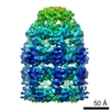 EMDB-1531: |
| Source |
|

 Escherichia coli (E. coli)
