 Movie
Movie Controller

[English] 日本語
 Yorodumi
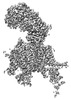
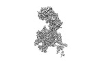
- PDB-8we6: Human L-type voltage-gated calcium channel Cav1.2 at 2.9 Angstrom... -
+ Open data

- Basic information
| Entry | Database: PDB / ID: 8we6 | ||||||
|---|---|---|---|---|---|---|---|
| Title | Human L-type voltage-gated calcium channel Cav1.2 at 2.9 Angstrom resolution | ||||||
 Components Components |
| ||||||
 Keywords Keywords |  MEMBRANE PROTEIN / Cav1.2 / Channels / Calcium Ion-Selective / TRANSPORT PROTEIN MEMBRANE PROTEIN / Cav1.2 / Channels / Calcium Ion-Selective / TRANSPORT PROTEIN | ||||||
| Function / homology |  Function and homology information: / voltage-gated calcium channel activity involved in AV node cell action potential / voltage-gated calcium channel activity involved in cardiac muscle cell action potential / immune system development / regulation of membrane repolarization during action potential / Presynaptic depolarization and calcium channel opening / positive regulation of high voltage-gated calcium channel activity / membrane depolarization during atrial cardiac muscle cell action potential / Phase 2 - plateau phase / calcium ion transmembrane transport via high voltage-gated calcium channel ...: / voltage-gated calcium channel activity involved in AV node cell action potential / voltage-gated calcium channel activity involved in cardiac muscle cell action potential / immune system development / regulation of membrane repolarization during action potential / Presynaptic depolarization and calcium channel opening / positive regulation of high voltage-gated calcium channel activity / membrane depolarization during atrial cardiac muscle cell action potential / Phase 2 - plateau phase / calcium ion transmembrane transport via high voltage-gated calcium channel / membrane depolarization during AV node cell action potential / membrane depolarization during bundle of His cell action potential / positive regulation of adenylate cyclase activity / cardiac conduction / L-type voltage-gated calcium channel complex / high voltage-gated calcium channel activity / membrane depolarization during cardiac muscle cell action potential / regulation of ventricular cardiac muscle cell action potential / cardiac muscle cell action potential involved in contraction / camera-type eye development / NCAM1 interactions / regulation of ventricular cardiac muscle cell membrane repolarization / embryonic forelimb morphogenesis / calcium ion transport into cytosol / cell communication by electrical coupling involved in cardiac conduction / regulation of calcium ion transmembrane transport via high voltage-gated calcium channel / voltage-gated calcium channel complex / neuromuscular junction development / neuronal dense core vesicle / Phase 0 - rapid depolarisation / regulation of heart rate by cardiac conduction / alpha-actinin binding / calcium channel regulator activity / regulation of calcium ion transport / calcium ion import across plasma membrane / voltage-gated calcium channel activity / regulation of cardiac muscle contraction by regulation of the release of sequestered calcium ion / sarcoplasmic reticulum / Regulation of insulin secretion / protein localization to plasma membrane / calcium ion transmembrane transport / postsynaptic density membrane / Z disc / Adrenaline,noradrenaline inhibits insulin secretion / cellular response to amyloid-beta / calcium ion transport / heart development / T cell receptor signaling pathway / positive regulation of cytosolic calcium ion concentration / perikaryon / chemical synaptic transmission / postsynaptic density / calmodulin binding / synapse / dendrite / extracellular exosome / membrane / metal ion binding / plasma membrane / cytosol / cytoplasm Function and homology information: / voltage-gated calcium channel activity involved in AV node cell action potential / voltage-gated calcium channel activity involved in cardiac muscle cell action potential / immune system development / regulation of membrane repolarization during action potential / Presynaptic depolarization and calcium channel opening / positive regulation of high voltage-gated calcium channel activity / membrane depolarization during atrial cardiac muscle cell action potential / Phase 2 - plateau phase / calcium ion transmembrane transport via high voltage-gated calcium channel ...: / voltage-gated calcium channel activity involved in AV node cell action potential / voltage-gated calcium channel activity involved in cardiac muscle cell action potential / immune system development / regulation of membrane repolarization during action potential / Presynaptic depolarization and calcium channel opening / positive regulation of high voltage-gated calcium channel activity / membrane depolarization during atrial cardiac muscle cell action potential / Phase 2 - plateau phase / calcium ion transmembrane transport via high voltage-gated calcium channel / membrane depolarization during AV node cell action potential / membrane depolarization during bundle of His cell action potential / positive regulation of adenylate cyclase activity / cardiac conduction / L-type voltage-gated calcium channel complex / high voltage-gated calcium channel activity / membrane depolarization during cardiac muscle cell action potential / regulation of ventricular cardiac muscle cell action potential / cardiac muscle cell action potential involved in contraction / camera-type eye development / NCAM1 interactions / regulation of ventricular cardiac muscle cell membrane repolarization / embryonic forelimb morphogenesis / calcium ion transport into cytosol / cell communication by electrical coupling involved in cardiac conduction / regulation of calcium ion transmembrane transport via high voltage-gated calcium channel / voltage-gated calcium channel complex / neuromuscular junction development / neuronal dense core vesicle / Phase 0 - rapid depolarisation / regulation of heart rate by cardiac conduction / alpha-actinin binding / calcium channel regulator activity / regulation of calcium ion transport / calcium ion import across plasma membrane / voltage-gated calcium channel activity / regulation of cardiac muscle contraction by regulation of the release of sequestered calcium ion / sarcoplasmic reticulum / Regulation of insulin secretion / protein localization to plasma membrane / calcium ion transmembrane transport / postsynaptic density membrane / Z disc / Adrenaline,noradrenaline inhibits insulin secretion / cellular response to amyloid-beta / calcium ion transport / heart development / T cell receptor signaling pathway / positive regulation of cytosolic calcium ion concentration / perikaryon / chemical synaptic transmission / postsynaptic density / calmodulin binding / synapse / dendrite / extracellular exosome / membrane / metal ion binding / plasma membrane / cytosol / cytoplasmSimilarity search - Function | ||||||
| Biological species |  Homo sapiens (human) Homo sapiens (human) | ||||||
| Method | ELECTRON MICROSCOPY / single particle reconstruction / cryo EM / Resolution: 2.9 Å | ||||||
 Authors Authors | Gao, S. / Yao, X. / Yan, N. | ||||||
| Funding support |  China, 1items China, 1items
| ||||||
 Citation Citation | Journal: Cell / Year: 2023 Title: Structural basis for human Ca1.2 inhibition by multiple drugs and the neurotoxin calciseptine. Authors: Shuai Gao / Xia Yao / Jiaofeng Chen / Gaoxingyu Huang / Xiao Fan / Lingfeng Xue / Zhangqiang Li / Tong Wu / Yupeng Zheng / Jian Huang / Xueqin Jin / Yan Wang / Zhifei Wang / Yong Yu / Lei ...Authors: Shuai Gao / Xia Yao / Jiaofeng Chen / Gaoxingyu Huang / Xiao Fan / Lingfeng Xue / Zhangqiang Li / Tong Wu / Yupeng Zheng / Jian Huang / Xueqin Jin / Yan Wang / Zhifei Wang / Yong Yu / Lei Liu / Xiaojing Pan / Chen Song / Nieng Yan /  Abstract: Ca1.2 channels play crucial roles in various neuronal and physiological processes. Here, we present cryo-EM structures of human Ca1.2, both in its apo form and in complex with several drugs, as well ...Ca1.2 channels play crucial roles in various neuronal and physiological processes. Here, we present cryo-EM structures of human Ca1.2, both in its apo form and in complex with several drugs, as well as the peptide neurotoxin calciseptine. Most structures, apo or bound to calciseptine, amlodipine, or a combination of amiodarone and sofosbuvir, exhibit a consistent inactivated conformation with a sealed gate, three up voltage-sensing domains (VSDs), and a down VSD. Calciseptine sits on the shoulder of the pore domain, away from the permeation path. In contrast, when pinaverium bromide, an antispasmodic drug, is inserted into a cavity reminiscent of the IFM-binding site in Na channels, a series of structural changes occur, including upward movement of VSD coupled with dilation of the selectivity filter and its surrounding segments in repeat III. Meanwhile, S4-5 merges with S5 to become a single helix, resulting in a widened but still non-conductive intracellular gate. | ||||||
| History |
|
- Structure visualization
| Structure viewer | Molecule: MolmilJmol/JSmol |
|---|
- Downloads & links
-Download
| PDBx/mmCIF format | 8we6.cif.gz | 552.9 KB | Display | PDBx/mmCIF format |
|---|---|---|---|---|
| PDB format | pdb8we6.ent.gz | 427.3 KB | Display | PDB format |
| PDBx/mmJSON format | 8we6.json.gz | Tree view | PDBx/mmJSON format | |
| Others |  Other downloads Other downloads |
-Validation report
| Arichive directory | https://data.pdbj.org/pub/pdb/validation_reports/we/8we6ftp://data.pdbj.org/pub/pdb/validation_reports/we/8we6 | HTTPS FTP |
|---|
-Related structure data
| Related structure data |  37472MC  8fhsC  8we7C 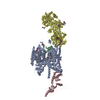 8we8C  8we9C  8weaC M: map data used to model this data C: citing same article ( |
|---|---|
| Similar structure data |


-Links
 PDBj
PDBj


- Assembly
| Deposited unit | 
|
|---|---|
| 1 |
|
-Components
-Voltage-dependent L-type calcium channel subunit ... , 2 types, 2 molecules AC
| #1: Protein | Mass: 246851.250 Da / Num. of mol.: 1 Source method: isolated from a genetically manipulated source Source: (gene. exp.) Homo sapiens (human) / Gene: CACNA1C / Production host: Homo sapiens (human) / References: UniProt: Q13936 |
|---|---|
| #3: Protein | Mass: 54607.852 Da / Num. of mol.: 1 Source method: isolated from a genetically manipulated source Source: (gene. exp.) Homo sapiens (human) / Gene: CACNB3 / Production host: Homo sapiens (human) / References: UniProt: P54284 |
-Protein , 1 types, 1 molecules D
| #2: Protein | Voltage-gated calcium channel / Voltage-gated calcium channel subunit alpha-2/delta-1 Mass: 124692.469 Da / Num. of mol.: 1 Source method: isolated from a genetically manipulated source Source: (gene. exp.) Homo sapiens (human) / Gene: CACNA2D1, CACNL2A, CCHL2A, MHS3 / Production host: Homo sapiens (human) / References: UniProt: P54289 |
|---|
-Sugars , 4 types, 8 molecules 
| #4: Polysaccharide | 2-acetamido-2-deoxy-beta-D-glucopyranose-(1-4)-2-acetamido-2-deoxy-beta-D-glucopyranose-(1-4)-2- ...2-acetamido-2-deoxy-beta-D-glucopyranose-(1-4)-2-acetamido-2-deoxy-beta-D-glucopyranose-(1-4)-2-acetamido-2-deoxy-beta-D-glucopyranose / Mass: 627.594 Da / Num. of mol.: 1 Source method: isolated from a genetically manipulated source | ||||
|---|---|---|---|---|---|
| #5: Polysaccharide | / Mass: 424.401 Da / Num. of mol.: 3 Source method: isolated from a genetically manipulated source #6: Polysaccharide | 2-acetamido-2-deoxy-beta-D-glucopyranose-(1-4)-2-acetamido-2-deoxy-beta-D-glucopyranose-(1-4)-2- ...2-acetamido-2-deoxy-beta-D-glucopyranose-(1-4)-2-acetamido-2-deoxy-beta-D-glucopyranose-(1-4)-2-acetamido-2-deoxy-beta-D-glucopyranose-(1-4)-2-acetamido-2-deoxy-beta-D-glucopyranose | / Mass: 830.786 Da / Num. of mol.: 1Source method: isolated from a genetically manipulated source #10: Sugar | N-Acetylglucosamine Type: D-saccharide, beta linking / Mass: 221.208 Da / Num. of mol.: 3 / Source method: obtained synthetically / Formula: C8H15NO6 Type: D-saccharide, beta linking / Mass: 221.208 Da / Num. of mol.: 3 / Source method: obtained synthetically / Formula: C8H15NO6 |
-Non-polymers , 4 types, 8 molecules 
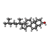
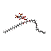
| #7: Chemical |  Mass: 40.078 Da / Num. of mol.: 2 / Source method: obtained synthetically / Formula: Ca Mass: 40.078 Da / Num. of mol.: 2 / Source method: obtained synthetically / Formula: Ca#8: Chemical | ChemComp-3PE / | Phosphatidylethanolamine Mass: 748.065 Da / Num. of mol.: 1 / Source method: obtained synthetically / Formula: C41H82NO8P / Comment: phospholipid*YM Mass: 748.065 Da / Num. of mol.: 1 / Source method: obtained synthetically / Formula: C41H82NO8P / Comment: phospholipid*YM#9: Chemical | ChemComp-CLR / Cholesterol Mass: 386.654 Da / Num. of mol.: 4 / Source method: obtained synthetically / Formula: C27H46O Mass: 386.654 Da / Num. of mol.: 4 / Source method: obtained synthetically / Formula: C27H46O#11: Chemical | ChemComp-PT5 / [( | Phosphatidylinositol 4,5-bisphosphate Mass: 1047.088 Da / Num. of mol.: 1 / Source method: obtained synthetically / Formula: C47H85O19P3 / Comment: phospholipid*YM Mass: 1047.088 Da / Num. of mol.: 1 / Source method: obtained synthetically / Formula: C47H85O19P3 / Comment: phospholipid*YM |
|---|
-Details
| Has ligand of interest | N |
|---|
-Experimental details
-Experiment
| Experiment | Method: ELECTRON MICROSCOPY |
|---|---|
| EM experiment | Aggregation state: PARTICLE / 3D reconstruction method: single particle reconstruction |
- Sample preparation
| Component | Name: Cav1.2 / Type: COMPLEX / Entity ID: #1-#3 / Source: RECOMBINANT |
|---|---|
| Molecular weight | Experimental value: NO |
| Source (natural) | Organism: Homo sapiens (human) |
| Source (recombinant) | Organism: Homo sapiens (human) |
| Buffer solution | pH: 7.4 |
| Specimen | Embedding applied: NO / Shadowing applied: NO / Staining applied: NO / Vitrification applied: YES |
| Vitrification | Cryogen name: ETHANE / Humidity: 100 % / Chamber temperature: 281 K |
- Electron microscopy imaging
| Experimental equipment |  Model: Titan Krios / Image courtesy: FEI Company |
|---|---|
| Microscopy | Model: FEI TITAN KRIOS |
| Electron gun | Electron source: FIELD EMISSION GUN / Accelerating voltage: 300 kV / Illumination mode: FLOOD BEAM |
| Electron lens | Mode: BRIGHT FIELDBright-field microscopy / Nominal defocus max: 2100 nm / Nominal defocus min: 1900 nm |
| Image recording | Electron dose: 50 e/Å2 / Film or detector model: GATAN K2 SUMMIT (4k x 4k) |
- Processing
| CTF correction | Type: PHASE FLIPPING AND AMPLITUDE CORRECTION | ||||||||||||||||||||||||
|---|---|---|---|---|---|---|---|---|---|---|---|---|---|---|---|---|---|---|---|---|---|---|---|---|---|
| 3D reconstruction | Resolution: 2.9 Å / Resolution method: FSC 0.143 CUT-OFF / Num. of particles: 74130 / Symmetry type: POINT | ||||||||||||||||||||||||
| Refine LS restraints |
|
