Journal: Curr Biol / Year: 2023 Title: Deep-branching evolutionary intermediates reveal structural origins of form I rubisco. Authors: Albert K Liu / Benjamin Kaeser / LinXing Chen / Jacob West-Roberts / Leah J Taylor-Kearney / Adi Lavy / Damian Günzing / Wen-Jun Li / Michal Hammel / Eva Nogales / Jillian F Banfield / Patrick M Shih / Abstract: The enzyme rubisco (ribulose-1,5-bisphosphate carboxylase/oxygenase) catalyzes the majority of biological carbon fixation on Earth. Although the vast majority of rubiscos across the tree of life ...The enzyme rubisco (ribulose-1,5-bisphosphate carboxylase/oxygenase) catalyzes the majority of biological carbon fixation on Earth. Although the vast majority of rubiscos across the tree of life assemble as homo-oligomers, the globally predominant form I enzyme-found in plants, algae, and cyanobacteria-forms a unique hetero-oligomeric complex. The recent discovery of a homo-oligomeric sister group to form I rubisco (named form I') has filled a key gap in our understanding of the enigmatic origins of the form I clade. However, to elucidate the series of molecular events leading to the evolution of form I rubisco, we must examine more distantly related sibling clades to contextualize the molecular features distinguishing form I and form I' rubiscos. Here, we present a comparative structural study retracing the evolutionary history of rubisco that reveals a complex structural trajectory leading to the ultimate hetero-oligomerization of the form I clade. We structurally characterize the oligomeric states of deep-branching form Iα and I'' rubiscos recently discovered from metagenomes, which represent key evolutionary intermediates preceding the form I clade. We further solve the structure of form I'' rubisco, revealing the molecular determinants that likely primed the enzyme core for the transition from a homo-oligomer to a hetero-oligomer. Our findings yield new insight into the evolutionary trajectory underpinning the adoption and entrenchment of the prevalent assembly of form I rubisco, providing additional context when viewing the enzyme family through the broader lens of protein evolution.
In the structure databanks used in Yorodumi, some data are registered as the other names, "COVID-19 virus" and "2019-nCoV". Here are the details of the virus and the list of structure data.
Jan 31, 2019. EMDB accession codes are about to change! (news from PDBe EMDB page)
EMDB accession codes are about to change! (news from PDBe EMDB page)
The allocation of 4 digits for EMDB accession codes will soon come to an end. Whilst these codes will remain in use, new EMDB accession codes will include an additional digit and will expand incrementally as the available range of codes is exhausted. The current 4-digit format prefixed with “EMD-” (i.e. EMD-XXXX) will advance to a 5-digit format (i.e. EMD-XXXXX), and so on. It is currently estimated that the 4-digit codes will be depleted around Spring 2019, at which point the 5-digit format will come into force.
The EM Navigator/Yorodumi systems omit the EMD- prefix.
Related info.:Q: What is EMD? / ID/Accession-code notation in Yorodumi/EM Navigator
Yorodumi is a browser for structure data from EMDB, PDB, SASBDB, etc.
This page is also the successor to EM Navigator detail page, and also detail information page/front-end page for Omokage search.
The word "yorodu" (or yorozu) is an old Japanese word meaning "ten thousand". "mi" (miru) is to see.
Related info.:EMDB / PDB / SASBDB / Comparison of 3 databanks / Yorodumi Search / Aug 31, 2016. New EM Navigator & Yorodumi / Yorodumi Papers / Jmol/JSmol / Function and homology information / Changes in new EM Navigator and Yorodumi
 Movie
Movie Controller


 Open data
 Basic information
 Components
 Keywords
 Function and homology information
 Bacillota (low GC Gram+)
 Authors
 United States, 2items
  Citation

 Structure visualization
 Downloads & links
 Other downloads
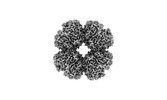
 PDBj
PDBj
 Assembly

 Mass: 24.305 Da / Num. of mol.: 8 / Source method: obtained synthetically / Formula: Mg / Feature type: SUBJECT OF INVESTIGATION
 Type: saccharide
 Mass: 18.015 Da / Num. of mol.: 981 / Source method: isolated from a natural source / Formula: H2O
 Sample preparation
 Electron microscopy imaging
 Processing