 ムービー
ムービー コントローラー


+ データを開く

- 基本情報
基本情報
| 登録情報 | データベース: PDB / ID: 8q6t | ||||||||||||||||||
|---|---|---|---|---|---|---|---|---|---|---|---|---|---|---|---|---|---|---|---|
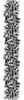
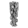
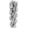
| タイトル | Helical reconstruction of the relaxed thick filament from FIB milled left ventricular mouse myofibrils | ||||||||||||||||||
 要素 要素 |
| ||||||||||||||||||
 キーワード キーワード |  MOTOR PROTEIN (モータータンパク質) / Mammalian (哺乳類) / Muscle (骨格筋) / Thick filament (ミオフィラメント) / Cardiac (心臓) MOTOR PROTEIN (モータータンパク質) / Mammalian (哺乳類) / Muscle (骨格筋) / Thick filament (ミオフィラメント) / Cardiac (心臓) | ||||||||||||||||||
| 機能・相同性 |  機能・相同性情報 機能・相同性情報forward locomotion / heart growth / striated muscle cell development / regulation of relaxation of cardiac muscle / muscle cell fate specification / Striated Muscle Contraction / regulation of slow-twitch skeletal muscle fiber contraction / regulation of the force of skeletal muscle contraction / sarcomerogenesis / structural molecule activity conferring elasticity ...forward locomotion / heart growth / striated muscle cell development / regulation of relaxation of cardiac muscle / muscle cell fate specification / Striated Muscle Contraction / regulation of slow-twitch skeletal muscle fiber contraction / regulation of the force of skeletal muscle contraction / sarcomerogenesis / structural molecule activity conferring elasticity / telethonin binding / contractile muscle fiber / skeletal muscle myosin thick filament assembly / cardiac myofibril / muscle myosin complex / regulation of striated muscle contraction / cardiac myofibril assembly / muscle filament sliding / muscle alpha-actinin binding / detection of muscle stretch / ventricular system development / regulation of the force of heart contraction / transition between fast and slow fiber / muscle cell development / myosin filament / cardiac muscle tissue morphogenesis / myosin II complex / adult heart development / cardiac muscle tissue development / cardiac muscle hypertrophy in response to stress / cardiac muscle hypertrophy / M band / actinin binding / I band / cardiac muscle cell development / myosin complex / A band / sarcomere organization / structural constituent of muscle / microfilament motor activity / ankyrin binding / ventricular cardiac muscle tissue morphogenesis / myofibril / heart contraction / intracellular non-membrane-bounded organelle / positive regulation of the force of heart contraction / skeletal muscle thin filament assembly / cytoskeletal motor activity / striated muscle thin filament / skeletal muscle contraction / actin monomer binding / striated muscle contraction / somitogenesis / ATP metabolic process / stress fiber / heart morphogenesis / protein kinase A signaling / cardiac muscle contraction / regulation of heart rate / sarcomere / post-embryonic development / muscle contraction / condensed nuclear chromosome / positive regulation of protein secretion / structural constituent of cytoskeleton / negative regulation of cell growth / Z disc / response to calcium ion / : / actin filament binding / マイクロフィラメント / heart development / protein tyrosine kinase activity / in utero embryonic development / protease binding / 細胞骨格 / calmodulin binding / non-specific serine/threonine protein kinase / リン酸化 / protein serine kinase activity / protein serine/threonine kinase activity / calcium ion binding / protein-containing complex binding / positive regulation of gene expression / protein kinase binding / enzyme binding / ATP hydrolysis activity / ATP binding / identical protein binding / 細胞質類似検索 - 分子機能 | ||||||||||||||||||
| 生物種 |  Mus musculus (ハツカネズミ) Mus musculus (ハツカネズミ) | ||||||||||||||||||
| 手法 | 電子顕微鏡法 / サブトモグラム平均法 / クライオ電子顕微鏡法 / 解像度: 18 Å | ||||||||||||||||||
 データ登録者 データ登録者 | Tamborrini, D. / Raunser, S. | ||||||||||||||||||
| 資金援助 |  ドイツ, European Union, ドイツ, European Union,  英国, 5件 英国, 5件
| ||||||||||||||||||
 引用 引用 | ジャーナル: Nature / 年: 2023 タイトル: Structure of the native myosin filament in the relaxed cardiac sarcomere. 著者: Davide Tamborrini / Zhexin Wang / Thorsten Wagner / Sebastian Tacke / Markus Stabrin / Michael Grange / Ay Lin Kho / Martin Rees / Pauline Bennett / Mathias Gautel / Stefan Raunser / 要旨: The thick filament is a key component of sarcomeres, the basic units of striated muscle. Alterations in thick filament proteins are associated with familial hypertrophic cardiomyopathy and other ...The thick filament is a key component of sarcomeres, the basic units of striated muscle. Alterations in thick filament proteins are associated with familial hypertrophic cardiomyopathy and other heart and muscle diseases. Despite the central importance of the thick filament, its molecular organization remains unclear. Here we present the molecular architecture of native cardiac sarcomeres in the relaxed state, determined by cryo-electron tomography. Our reconstruction of the thick filament reveals the three-dimensional organization of myosin, titin and myosin-binding protein C (MyBP-C). The arrangement of myosin molecules is dependent on their position along the filament, suggesting specialized capacities in terms of strain susceptibility and force generation. Three pairs of titin-α and titin-β chains run axially along the filament, intertwining with myosin tails and probably orchestrating the length-dependent activation of the sarcomere. Notably, whereas the three titin-α chains run along the entire length of the thick filament, titin-β chains do not. The structure also demonstrates that MyBP-C bridges thin and thick filaments, with its carboxy-terminal region binding to the myosin tails and directly stabilizing the OFF state of the myosin heads in an unforeseen manner. These results provide a foundation for future research investigating muscle disorders involving sarcomeric components. | ||||||||||||||||||
| 履歴 |
|
- 構造の表示
構造の表示
| 構造ビューア | 分子: MolmilJmol/JSmol |
|---|
- ダウンロードとリンク
-ダウンロード
| PDBx/mmCIF形式 | 8q6t.cif.gz | 2.7 MB | 表示 | PDBx/mmCIF形式 |
|---|---|---|---|---|
| PDB形式 | pdb8q6t.ent.gz | 表示 | PDB形式 | |
| PDBx/mmJSON形式 | 8q6t.json.gz | ツリー表示 | PDBx/mmJSON形式 | |
| その他 |  その他のダウンロード その他のダウンロード |
-検証レポート
| アーカイブディレクトリ | https://data.pdbj.org/pub/pdb/validation_reports/q6/8q6tftp://data.pdbj.org/pub/pdb/validation_reports/q6/8q6t | HTTPS FTP |
|---|
-関連構造データ


-リンク
 PDBj
PDBj


- 集合体
集合体
| 登録構造単位 | 
|
|---|---|
| 1 |
|
-要素
| #1: タンパク質 | ミオシン / Myosin heavy chain 7 / Myosin heavy chain slow isoform / MyHC-slow / Myosin heavy chain / cardiac ...Myosin heavy chain 7 / Myosin heavy chain slow isoform / MyHC-slow / Myosin heavy chain / cardiac muscle beta isoform / MyHC-beta 分子量: 223226.531 Da / 分子数: 6 / 由来タイプ: 天然 / 由来: (天然) Mus musculus (ハツカネズミ) / 参照: UniProt: Q91Z83#2: タンパク質 | 分子量: 17243.553 Da / 分子数: 6 / 由来タイプ: 天然 / 由来: (天然) Mus musculus (ハツカネズミ) / 参照: UniProt: P09542#3: タンパク質 | 分子量: 18259.512 Da / 分子数: 6 / 由来タイプ: 天然 / 由来: (天然) Mus musculus (ハツカネズミ) / 参照: UniProt: P51667#4: タンパク質 | 分子量: 44777.125 Da / 分子数: 2 / 由来タイプ: 天然 / 由来: (天然) Mus musculus (ハツカネズミ) / 参照: UniProt: Q3TF37#5: タンパク質 | チチン分子量: 118766.320 Da / 分子数: 2 / 由来タイプ: 天然 / 由来: (天然) Mus musculus (ハツカネズミ) / 参照: UniProt: A2ASS6 |
|---|
-実験情報
-実験
| 実験 | 手法: 電子顕微鏡法 |
|---|---|
| EM実験 | 試料の集合状態: CELL / 3次元再構成法: サブトモグラム平均法 |
- 試料調製
試料調製
| 構成要素 | 名称: Relaxed thick filament; A-band region; C-type super-repeat タイプ: CELL 詳細: Single asymmetrical unit from the relaxed thick filament obtained from FIB milled left ventricular mouse myofibrils Entity ID: #1-#3, #5, #4 / 由来: NATURAL |
|---|---|
| 由来(天然) | 生物種: Mus musculus (ハツカネズミ) |
| 緩衝液 | pH: 7.1 |
| 試料 | 包埋: NO / シャドウイング: NO / 染色: NO / 凍結: YES |
| 急速凍結 | 凍結剤: ETHANE-PROPANE |
- 電子顕微鏡撮影
| 実験機器 |  モデル: Titan Krios / 画像提供: FEI Company |
|---|---|
| 顕微鏡 | モデル: FEI TITAN KRIOS |
| 電子銃 | 電子線源: FIELD EMISSION GUN / 加速電圧: 300 kV / 照射モード: FLOOD BEAM |
| 電子レンズ | モード: BRIGHT FIELDBright-field microscopy / 倍率(公称値): 81000 X / 最大 デフォーカス(公称値): 6000 nm / 最小 デフォーカス(公称値): 3000 nm |
| 試料ホルダ | 試料ホルダーモデル: FEI TITAN KRIOS AUTOGRID HOLDER |
| 撮影 | 電子線照射量: 3.4 e/Å2 / Avg electron dose per subtomogram: 140 e/Å2 フィルム・検出器のモデル: GATAN K3 BIOQUANTUM (6k x 4k) |
- 解析
解析
| EMソフトウェア |
| |||||||||||||||||||||
|---|---|---|---|---|---|---|---|---|---|---|---|---|---|---|---|---|---|---|---|---|---|---|
| CTF補正 | タイプ: PHASE FLIPPING AND AMPLITUDE CORRECTION | |||||||||||||||||||||
| らせん対称 | 回転角度/サブユニット: 0 ° / 軸方向距離/サブユニット: 430 Å / らせん対称軸の対称性: C3 | |||||||||||||||||||||
| 3次元再構成 | 解像度: 18 Å / 解像度の算出法: FSC 0.143 CUT-OFF / 粒子像の数: 1589 詳細: Helical reconstruction containing 4.5x repeats extrapolated from a 3x repeat reconstruction (EMD-18146) 対称性のタイプ: HELICAL | |||||||||||||||||||||
| EM volume selection | Num. of tomograms: 89 / Num. of volumes extracted: 67492 | |||||||||||||||||||||
| 原子モデル構築 |
|
