 Movie
Movie Controller

[English] 日本語
 Yorodumi
- EMDB-18198: Helical reconstruction of the relaxed thick filament from FIB mil... -
+ Open data

- Basic information
| Entry | 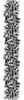 | ||||||||||||||||||
|---|---|---|---|---|---|---|---|---|---|---|---|---|---|---|---|---|---|---|---|
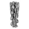
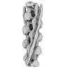
| Title | Helical reconstruction of the relaxed thick filament from FIB milled left ventricular mouse myofibrils | ||||||||||||||||||
 Map data Map data | Helical reconstruction of the relaxed thick filament from FIB-milled mouse left ventricular myofibrils | ||||||||||||||||||
 Sample Sample |
| ||||||||||||||||||
 Keywords Keywords |  Mammalian / Muscle / Thick filament / Cardiac / MOTOR PROTEIN Mammalian / Muscle / Thick filament / Cardiac / MOTOR PROTEIN | ||||||||||||||||||
| Function / homology |  Function and homology information Function and homology informationforward locomotion / heart growth / striated muscle cell development / regulation of relaxation of cardiac muscle / muscle cell fate specification / Striated Muscle Contraction / regulation of slow-twitch skeletal muscle fiber contraction / regulation of the force of skeletal muscle contraction / sarcomerogenesis / structural molecule activity conferring elasticity ...forward locomotion / heart growth / striated muscle cell development / regulation of relaxation of cardiac muscle / muscle cell fate specification / Striated Muscle Contraction / regulation of slow-twitch skeletal muscle fiber contraction / regulation of the force of skeletal muscle contraction / sarcomerogenesis / structural molecule activity conferring elasticity / telethonin binding / contractile muscle fiber / skeletal muscle myosin thick filament assembly / cardiac myofibril / muscle myosin complex / regulation of striated muscle contraction / cardiac myofibril assembly / muscle filament sliding / muscle alpha-actinin binding / detection of muscle stretch / ventricular system development / regulation of the force of heart contraction / transition between fast and slow fiber / muscle cell development / myosin filament / cardiac muscle tissue morphogenesis / myosin II complex / adult heart development / cardiac muscle tissue development / cardiac muscle hypertrophy in response to stress / cardiac muscle hypertrophy / M band / actinin binding / I band / cardiac muscle cell development / myosin complex / A band / sarcomere organization / structural constituent of muscle / microfilament motor activity / ankyrin binding / ventricular cardiac muscle tissue morphogenesis / myofibril / heart contraction / intracellular non-membrane-bounded organelle / positive regulation of the force of heart contraction / skeletal muscle thin filament assembly / cytoskeletal motor activity / striated muscle thin filament / skeletal muscle contraction / actin monomer binding / striated muscle contraction / somitogenesis / ATP metabolic process / stress fiber / heart morphogenesis / protein kinase A signaling / cardiac muscle contraction / regulation of heart rate / sarcomere / post-embryonic development / muscle contraction / condensed nuclear chromosome / positive regulation of protein secretion / structural constituent of cytoskeleton / negative regulation of cell growth / Z disc / response to calcium ion / : / actin filament binding / actin cytoskeleton / heart development / protein tyrosine kinase activity / in utero embryonic development / protease binding / cytoskeleton / calmodulin binding / non-specific serine/threonine protein kinase / phosphorylation / protein serine kinase activity / protein serine/threonine kinase activity / calcium ion binding / protein-containing complex binding / positive regulation of gene expression / protein kinase binding / enzyme binding / ATP hydrolysis activity / ATP binding / identical protein binding / cytoplasmSimilarity search - Function | ||||||||||||||||||
| Biological species |  Mus musculus (house mouse) Mus musculus (house mouse) | ||||||||||||||||||
| Method | subtomogram averaging / cryo EM / Resolution: 18.0 Å | ||||||||||||||||||
 Authors Authors | Tamborrini D / Raunser S | ||||||||||||||||||
| Funding support |  Germany, European Union, Germany, European Union,  United Kingdom, 5 items United Kingdom, 5 items
| ||||||||||||||||||
 Citation Citation | Journal: Nature / Year: 2023 Title: Structure of the native myosin filament in the relaxed cardiac sarcomere. Authors: Davide Tamborrini / Zhexin Wang / Thorsten Wagner / Sebastian Tacke / Markus Stabrin / Michael Grange / Ay Lin Kho / Martin Rees / Pauline Bennett / Mathias Gautel / Stefan Raunser / Abstract: The thick filament is a key component of sarcomeres, the basic units of striated muscle. Alterations in thick filament proteins are associated with familial hypertrophic cardiomyopathy and other ...The thick filament is a key component of sarcomeres, the basic units of striated muscle. Alterations in thick filament proteins are associated with familial hypertrophic cardiomyopathy and other heart and muscle diseases. Despite the central importance of the thick filament, its molecular organization remains unclear. Here we present the molecular architecture of native cardiac sarcomeres in the relaxed state, determined by cryo-electron tomography. Our reconstruction of the thick filament reveals the three-dimensional organization of myosin, titin and myosin-binding protein C (MyBP-C). The arrangement of myosin molecules is dependent on their position along the filament, suggesting specialized capacities in terms of strain susceptibility and force generation. Three pairs of titin-α and titin-β chains run axially along the filament, intertwining with myosin tails and probably orchestrating the length-dependent activation of the sarcomere. Notably, whereas the three titin-α chains run along the entire length of the thick filament, titin-β chains do not. The structure also demonstrates that MyBP-C bridges thin and thick filaments, with its carboxy-terminal region binding to the myosin tails and directly stabilizing the OFF state of the myosin heads in an unforeseen manner. These results provide a foundation for future research investigating muscle disorders involving sarcomeric components. | ||||||||||||||||||
| History |
|
- Structure visualization
| Supplemental images |
|---|

- Downloads & links
-EMDB archive
| Map data | emd_18198.map.gz | 1.9 MB | EMDB map data format | |
|---|---|---|---|---|
| Header (meta data) | emd-18198-v30.xmlemd-18198.xml | 19.5 KB 19.5 KB | Display Display | EMDB header |
| Images |  emd_18198.png emd_18198.png | 43 KB | ||
| Filedesc metadata | emd-18198.cif.gz | 8.1 KB | ||
| Archive directory |  http://ftp.pdbj.org/pub/emdb/structures/EMD-18198ftp://ftp.pdbj.org/pub/emdb/structures/EMD-18198 http://ftp.pdbj.org/pub/emdb/structures/EMD-18198ftp://ftp.pdbj.org/pub/emdb/structures/EMD-18198 | HTTPS FTP |
-Related structure data
| Related structure data |  8q6tMC  8q4gC C: citing same article ( M: atomic model generated by this map |
|---|---|
| Similar structure data |


-Links
| EMDB pages | EMDB (EBI/PDBe) / EMDataResource |
|---|---|
| Related items in Molecule of the Month |


-Map
| File | Download / File: emd_18198.map.gz / Format: CCP4 / Size: 196.4 MB / Type: IMAGE STORED AS FLOATING POINT NUMBER (4 BYTES) | ||||||||||||||||||||
|---|---|---|---|---|---|---|---|---|---|---|---|---|---|---|---|---|---|---|---|---|---|
| Annotation | Helical reconstruction of the relaxed thick filament from FIB-milled mouse left ventricular myofibrils | ||||||||||||||||||||
| Voxel size | X=Y=Z: 5.83 Å | ||||||||||||||||||||
| Density |
| ||||||||||||||||||||
| Symmetry | Space group: 1 | ||||||||||||||||||||
| Details | EMDB XML:
|
-Supplemental data
- Sample components
-Entire : Relaxed thick filament; A-band region; C-type super-repeat
| Entire | Name: Relaxed thick filament; A-band region; C-type super-repeat |
|---|---|
| Components |
|
-Supramolecule #1: Relaxed thick filament; A-band region; C-type super-repeat
| Supramolecule | Name: Relaxed thick filament; A-band region; C-type super-repeat type: cell / ID: 1 / Parent: 0 / Macromolecule list: #1-#3, #5, #4 Details: Single asymmetrical unit from the relaxed thick filament obtained from FIB milled left ventricular mouse myofibrils |
|---|---|
| Source (natural) | Organism: Mus musculus (house mouse) |
-Macromolecule #1: Myosin-7
| Macromolecule | Name: Myosin-7 / type: protein_or_peptide / ID: 1 / Number of copies: 6 / Enantiomer: LEVO |
|---|---|
| Source (natural) | Organism: Mus musculus (house mouse) |
| Molecular weight | Theoretical: 223.226531 KDa |
| Sequence | String: MADAEMAAFG AAAPFLRKSE KERLEAQTRP FDLKKDVFVP DDKEEFVKAK IVSREGGKVT AETENGKTVT VKEDQVMQQN PPKFDKIED MAMLTFLHEP AVLYNLKERY ASWMIYTYSG LFCVTVNPYK WLPVYNAEVV AAYRGKKRSE APPHIFSISD N AYQYMLTD ...String: MADAEMAAFG AAAPFLRKSE KERLEAQTRP FDLKKDVFVP DDKEEFVKAK IVSREGGKVT AETENGKTVT VKEDQVMQQN PPKFDKIED MAMLTFLHEP AVLYNLKERY ASWMIYTYSG LFCVTVNPYK WLPVYNAEVV AAYRGKKRSE APPHIFSISD N AYQYMLTD RENQSILITG ESGAGKTVNT KRVIQYFAVI AAIGDRSKKD QTPGKGTLED QIIQANPALE AFGNAKTVRN DN SSRFGKF IRIHFGATGK LASADIETYL LEKSRVIFQL KAERDYHIFY QILSNKKPEL LDMLLITNNP YDYAFISQGE TTV ASIDDS EELMATDSAF DVLGFTPEEK NSIYKLTGAI MHFGNMKFKQ KQREEQAEPD GTEEADKSAY LMGLNSADLL KGLC HPRVK VGNEYVTKGQ NVQQVSYAIG ALAKSVYEKM FNWMVTRINA TLETKQPRQY FIGVLDIAGF EIFDFNSFEQ LCINF TNEK LQQFFNHHMF VLEQEEYKKE GIEWTFIDFG MDLQACIDLI EKPMGIMSIL EEECMFPKAT DMTFKAKLYD NHLGKS NNF QKPRNVKGKQ EAHFSLVHYA GTVDYNILGW LQKNKDPLNE TVVGLYQKSS LKLLSNLFAN YAGADAPADK GKGKAKK GS SFQTVSALHR ENLNKLMTNL RSTHPHFVRC IIPNETKSPG VMDNPLVMHQ LRCNGVLEGI RICRKGFPNR ILYGDFRQ R YRILNPAAIP EGQFIDSRKG AEKLLGSLDI DHNQYKFGHT KVFFKAGLLG LLEEMRDERL SRIITRIQAQ SRGVLSRME FKKLLERRDS LLIIQWNIRA FMGVKNWPWM KLYFKIKPLL KSAETEKEMA TMKEEFGRVK DALEKSEARR KELEEKMVSL LQEKNDLQL QVQAEQDNLA DAEERCDQLI KNKIQLEAKV KEMTERLEDE EEMNAELTAK KRKLEDECSE LKRDIDDLEL T LAKVEKEK HATENKVKNL TEEMAGLDEI IVKLTKEKKA LQEAHQQALD DLQAEEDKVN TLTKAKVKLE QQVDDLEGSL EQ EKKVRMD LERAKRKLEG DLKLTQESIM DLENDKQQLD ERLKKKDFEL NALNARIEDE QALGSQLQKK LKELQARIEE LEE ELEAER TARAKVEKLR SDLSRELEEI SERLEEAGGA TSVQIEMNKK REAEFQKMRR DLEEATLQHE ATAAALRKKH ADSV AELGE QIDNLQRVKQ KLEKEKSEFK LELDDVTSNM EQIIKAKANL EKMCRTLEDQ MNEHRSKAEE TQRSVNDLTS QRAKL QTEN GELSRQLDEK EALISQLTRG KLTYTQQLED LKRQLEEEVK AKNALAHALQ SARHDCDLLR EQYEEETEAK AELQRV LSK ANSEVAQWRT KYETDAIQRT EELEEAKKKL AQRLQDAEEA VEAVNAKCSS LEKTKHRLQN EIEDLMVDVE RSNAAAA AL DKKQRNFDKI LAEWKQKYEE SQSELESSQK EARSLSTELF KLKNAYEESL EHLETFKREN KNLQEEISDL TEQLGSTG K SIHELEKIRK QLEAEKLELQ SALEEAEASL EHEEGKILRA QLEFNQIKAE IERKLAEKDE EMEQAKRNHL RMVDSLQTS LDAETRSRNE ALRVKKKMEG DLNEMEIQLS HANRMAAEAQ KQVKSLQSLL KDTQIQLDDA VRANDDLKEN IAIVERRNNL LQAELEELR AVVEQTERSR KLAEQELIET SERVQLLHSQ NTSLINQKKK MDADLSQLQT EVEEAVQECR NAEEKAKKAI T DAAMMAEE LKKEQDTSAH LERMKKNMEQ TIKDLQHRLD EAEQIALKGG KKQLQKLEAR VRELENELEA EQKRNAESVK GM RKSERRI KELTYQTEED RKNLLRLQDL VDKLQLKVKA YKRQAEEAEE QANTNLSKFR KVQHELDEAE ERADIAESQV NKL RAKSRD IGAKGLNEE UniProtKB: Myosin-7 |
-Macromolecule #2: Myosin light chain 3
| Macromolecule | Name: Myosin light chain 3 / type: protein_or_peptide / ID: 2 / Number of copies: 6 / Enantiomer: LEVO |
|---|---|
| Source (natural) | Organism: Mus musculus (house mouse) |
| Molecular weight | Theoretical: 17.243553 KDa |
| Sequence | String: IEFTPEQIEE FKEAFLLFDR TPKGEMKITY GQCGDVLRAL GQNPTQAEVL RVLGKPKQEE LNSKMMDFET FLPMLQHISK NKDTGTYED FVEGLRVFDK EGNGTVMGAE LRHVLATLGE RLTEDEVEKL MAGQEDSNGC INYEAFVKHI MAS UniProtKB: Myosin light chain 3 |
-Macromolecule #3: Myosin regulatory light chain 2, ventricular/cardiac muscle isoform
| Macromolecule | Name: Myosin regulatory light chain 2, ventricular/cardiac muscle isoform type: protein_or_peptide / ID: 3 / Number of copies: 6 / Enantiomer: LEVO |
|---|---|
| Source (natural) | Organism: Mus musculus (house mouse) |
| Molecular weight | Theoretical: 18.259512 KDa |
| Sequence | String: KKRIEGGSSN VFSMFEQTQI QEFKEAFTIM DQNRDGFIDK NDLRDTFAAL GRVNVKNEEI DEMIKEAPGP INFTVFLTMF GEKLKGADP EETILNAFKV FDPEGKGSLK ADYVREMLTT QAERFSKEEI DQMFAAFPPD VTGNLDYKNL VHIITHGEEK D UniProtKB: Myosin regulatory light chain 2, ventricular/cardiac muscle isoform |
-Macromolecule #4: Myosin binding protein C, cardiac
| Macromolecule | Name: Myosin binding protein C, cardiac / type: protein_or_peptide / ID: 4 / Number of copies: 2 / Enantiomer: LEVO |
|---|---|
| Source (natural) | Organism: Mus musculus (house mouse) |
| Molecular weight | Theoretical: 44.777125 KDa |
| Sequence | String: PIGPPGEPTH LAVEDVSDTT VSLKWRPPER VGAGGLDGYS VEYCQEGCSE WTPALQGLTE RTSMLVKDLP TGARLLFRVR AHNVAGPGG PIVTKEPVTV QEILQRPRLQ LPRHLRQTIQ KKVGEPVNLL IPFQGKPRPQ VTWTKEGQPL AGEEVSIRNS P TDTILFIR ...String: PIGPPGEPTH LAVEDVSDTT VSLKWRPPER VGAGGLDGYS VEYCQEGCSE WTPALQGLTE RTSMLVKDLP TGARLLFRVR AHNVAGPGG PIVTKEPVTV QEILQRPRLQ LPRHLRQTIQ KKVGEPVNLL IPFQGKPRPQ VTWTKEGQPL AGEEVSIRNS P TDTILFIR AARRTHSGTY QVTVRIENME DKATLILQIV DKPSPPQDIR IVETWGFNVA LEWKPPQDDG NTEIWGYTVQ KA DKKTMEW FTVLEHYRRT HCVVSELIIG NGYYFRVFSH NMVGSSDKAA ATKEPVFIPR PGITYEPPKY KALDFSEAPS FTQ PLANRS IIAGYNAILC CAVRGSPKPK ISWFKNGLDL GEDARFRMFC KQGVLTLEIR KPCPYDGGVY VCRATNLQGE AQCE UniProtKB: Myosin binding protein C, cardiac |
-Macromolecule #5: Titin
| Macromolecule | Name: Titin / type: protein_or_peptide / ID: 5 / Number of copies: 2 / Enantiomer: LEVO |
|---|---|
| Source (natural) | Organism: Mus musculus (house mouse) |
| Molecular weight | Theoretical: 118.76632 KDa |
| Sequence | String: DPCDPPGRPE AIVITRNSVT LKWKKPVYDG GSKITGYIVE KKDLPDGRWM KASFTNVVET EFTVTGLVED QRYEFRVIAR NAADNFSEP SESSGAITAR DEIDAPNASL DPKYRDVIIV HAGETFVLEA DIRGKPIPDI IWSKDGNELE ETAARMEIKS T LQKTTLIV ...String: DPCDPPGRPE AIVITRNSVT LKWKKPVYDG GSKITGYIVE KKDLPDGRWM KASFTNVVET EFTVTGLVED QRYEFRVIAR NAADNFSEP SESSGAITAR DEIDAPNASL DPKYRDVIIV HAGETFVLEA DIRGKPIPDI IWSKDGNELE ETAARMEIKS T LQKTTLIV KDCIRTDGGQ YTLKLSNVGG TKTIPITVKV LDRPGPPEGP LKVTGVTAEK CYLAWNPPLQ DGGASISHYI IE KRETSRL SWTQVSNEVQ ALNYKVTKLL PGNEYIFRVM AVNKYGIGEA LESEPVIACN PYKRPGPPST PEASAITKDS MVL TWTRPV DDGGAEIEGY ILEKRDKEGI RWTKCNKKTL TDLRFRVTGL TEGHSYEFRV AAENAAGVGE PSEPSVFYRA CDAL YPPGP PSNPKVTDTS RSSVSLAWNK PIYDGGAPVR GYVIELKKAA ADEWTTCTPP SGLQGKQFTV TKLKENTEYN FRICA FNTE GVGEPATIPG SVVAQERMEA PEIELDADLR KVVTLRASAT LRLFVTIKGR PEPEVKWEKA EGILTERAQI EVTSSY TML VIDNVTRFDS GRYNLTLENN SGSKTAFVNV RVLDSPSAPV NLTIREVKKD SVTLSWEPPL IDGGAKITNY IVEKRET TR KAYATITNNC TKNTFKIENL QEGCSYYFRV LASNEYGIGL PAETAEPVKV SEPPLPPGRV TLVDVTRNTA TIKWEKPE S DGGSKITGYV VEMQTKGSEK WSACTQVKTL ETTISGLTAG EEYVFRVAAV NEKGRSDPRQ LGVPVIAKDI EIKPSVELP FNTFNVKAND QLKIDIPFKG RPQATVAWKK DGQVLRETTR VNVASSKTVT TLSIKEASRE DVGTYELCVS NTAGSITVPI TVIVLDRPG PPGPIRIDEV SCDNVSISWN PPEYDGGCQI SNYIVEKRET TSTTWQVVSQ AVARTSIKIV RLTTGSEYQF R VCAENRYG KSSYSESSAV VAEYPFSPPG PPGTPKVVHA TKSTMVVSWQ VPVNDGGSQV IGYHLEYKER SSILWSKANK VL IADTQMK VSGLDEGLMY EYRVYAENIA GIGKCSKACE PV UniProtKB: Titin |
-Experimental details
-Structure determination
| Method | cryo EM |
|---|---|
 Processing Processing | subtomogram averaging |
| Aggregation state | cell |
-Sample preparation
| Buffer | pH: 7.1 |
|---|---|
| Vitrification | Cryogen name: ETHANE-PROPANE |
- Electron microscopy
| Microscope | FEI TITAN KRIOS |
|---|---|
| Electron beam | Acceleration voltage: 300 kV / Electron source: FIELD EMISSION GUN |
| Electron optics | Illumination mode: FLOOD BEAM / Imaging mode: BRIGHT FIELDBright-field microscopy / Nominal defocus max: 6.0 µm / Nominal defocus min: 3.0 µm / Nominal magnification: 81000 |
| Sample stage | Specimen holder model: FEI TITAN KRIOS AUTOGRID HOLDER |
| Image recording | Film or detector model: GATAN K3 BIOQUANTUM (6k x 4k) / Average electron dose: 3.4 e/Å2 |
| Experimental equipment |  Model: Titan Krios / Image courtesy: FEI Company |
-Image processing
| Extraction | Number tomograms: 89 / Number images used: 67492 |
|---|---|
| Final angle assignment | Type: MAXIMUM LIKELIHOOD / Software - Name: RELION |
| Final reconstruction | Applied symmetry - Helical parameters - Δz: 430.0 Å Applied symmetry - Helical parameters - Δ&Phi: 0 ° Applied symmetry - Helical parameters - Axial symmetry: C3 (3 fold cyclic )Resolution.type: BY AUTHOR / Resolution: 18.0 Å / Resolution method: FSC 0.143 CUT-OFF / Software - Name: RELION Details: Helical reconstruction containing 4.5x repeats extrapolated from a 3x repeat reconstruction (EMD-18146) Number subtomograms used: 1589 |
-Atomic model buiding 1
| Initial model | (Chain: AlphaFold, SwissModel) |
|---|---|
| Output model | PDB-8q6t: |
