 Movie
Movie Controller


+ Open data

- Basic information
| Entry | Database: PDB / ID: 8emt | ||||||
|---|---|---|---|---|---|---|---|
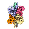
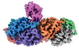
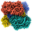
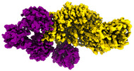
| Title | Cryo-EM analysis of the human aldehyde oxidase from liver | ||||||
 Components Components | Aldehyde oxidase | ||||||
 Keywords Keywords | OXIDOREDUCTASE / Aldehyde oxidase / AOX1 | ||||||
| Function / homology |  Function and homology informationOxidoreductases; Acting on CH or CH2 groups; With oxygen as acceptor / aldehyde oxidase / aldehyde oxidase activity / Vitamin B6 activation to pyridoxal phosphate / molybdopterin cofactor binding / xenobiotic metabolic process / FAD binding / lipid metabolic process / 2 iron, 2 sulfur cluster binding / NAD binding ...Oxidoreductases; Acting on CH or CH2 groups; With oxygen as acceptor / aldehyde oxidase / aldehyde oxidase activity / Vitamin B6 activation to pyridoxal phosphate / molybdopterin cofactor binding / xenobiotic metabolic process / FAD binding / lipid metabolic process / 2 iron, 2 sulfur cluster binding / NAD binding / flavin adenine dinucleotide binding / iron ion binding / protein homodimerization activity / extracellular exosome / identical protein binding / cytosol Function and homology informationOxidoreductases; Acting on CH or CH2 groups; With oxygen as acceptor / aldehyde oxidase / aldehyde oxidase activity / Vitamin B6 activation to pyridoxal phosphate / molybdopterin cofactor binding / xenobiotic metabolic process / FAD binding / lipid metabolic process / 2 iron, 2 sulfur cluster binding / NAD binding ...Oxidoreductases; Acting on CH or CH2 groups; With oxygen as acceptor / aldehyde oxidase / aldehyde oxidase activity / Vitamin B6 activation to pyridoxal phosphate / molybdopterin cofactor binding / xenobiotic metabolic process / FAD binding / lipid metabolic process / 2 iron, 2 sulfur cluster binding / NAD binding / flavin adenine dinucleotide binding / iron ion binding / protein homodimerization activity / extracellular exosome / identical protein binding / cytosolSimilarity search - Function | ||||||
| Biological species |  Homo sapiens (human) Homo sapiens (human) | ||||||
| Method | ELECTRON MICROSCOPY / single particle reconstruction / cryo EM / Resolution: 2.92 Å | ||||||
 Authors Authors | Su, C. / Lyu, M. / Zhang, Z. / Yu, E.W. | ||||||
| Funding support |  United States, 1items United States, 1items
| ||||||
 Citation Citation | Journal: Cell Rep / Year: 2023 Title: High-resolution structural-omics of human liver enzymes. Authors: Chih-Chia Su / Meinan Lyu / Zhemin Zhang / Masaru Miyagi / Wei Huang / Derek J Taylor / Edward W Yu / Abstract: We applied raw human liver microsome lysate to a holey carbon grid and used cryo-electron microscopy (cryo-EM) to define its composition. From this sample we identified and simultaneously determined ...We applied raw human liver microsome lysate to a holey carbon grid and used cryo-electron microscopy (cryo-EM) to define its composition. From this sample we identified and simultaneously determined high-resolution structural information for ten unique human liver enzymes involved in diverse cellular processes. Notably, we determined the structure of the endoplasmic bifunctional protein H6PD, where the N- and C-terminal domains independently possess glucose-6-phosphate dehydrogenase and 6-phosphogluconolactonase enzymatic activity, respectively. We also obtained the structure of heterodimeric human GANAB, an ER glycoprotein quality-control machinery that contains a catalytic α subunit and a noncatalytic β subunit. In addition, we observed a decameric peroxidase, PRDX4, which directly contacts a disulfide isomerase-related protein, ERp46. Structural data suggest that several glycosylations, bound endogenous compounds, and ions associate with these human liver enzymes. These results highlight the importance of cryo-EM in facilitating the elucidation of human organ proteomics at the atomic level. | ||||||
| History |
|
- Structure visualization
| Structure viewer | Molecule: MolmilJmol/JSmol |
|---|
- Downloads & links
-Download
| PDBx/mmCIF format | 8emt.cif.gz | 435.4 KB | Display | PDBx/mmCIF format |
|---|---|---|---|---|
| PDB format | pdb8emt.ent.gz | 350.8 KB | Display | PDB format |
| PDBx/mmJSON format | 8emt.json.gz | Tree view | PDBx/mmJSON format | |
| Others |  Other downloads Other downloads |
-Validation report
| Arichive directory | https://data.pdbj.org/pub/pdb/validation_reports/em/8emtftp://data.pdbj.org/pub/pdb/validation_reports/em/8emt | HTTPS FTP |
|---|
-Related structure data
| Related structure data |  28264MC 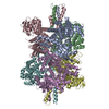 7uzmC  8ekwC  8ekyC  8em2C  8emrC  8emsC  8eneC  8eojC  8eorC M: map data used to model this data C: citing same article ( |
|---|---|
| Similar structure data | |
| Experimental dataset #1 | Data reference: 10.6019/EMPIAR-11250 / Data set type: EMPIAR |


-Links
 PDBj
PDBj


- Assembly
| Deposited unit | 
|
|---|---|
| 1 |
|
-Components
| #1: Protein | / Aldehyde oxidase 1 / Azaheterocycle hydroxylase Mass: 148096.500 Da / Num. of mol.: 2 / Source method: isolated from a natural source / Source: (natural) Homo sapiens (human)References: UniProt: Q06278, aldehyde oxidase, Oxidoreductases; Acting on CH or CH2 groups; With oxygen as acceptor#2: Chemical | ChemComp-FES / Iron–sulfur cluster  Mass: 175.820 Da / Num. of mol.: 4 / Source method: isolated from a natural source / Formula: Fe2S2 Mass: 175.820 Da / Num. of mol.: 4 / Source method: isolated from a natural source / Formula: Fe2S2#3: Chemical |   Mass: 395.352 Da / Num. of mol.: 2 / Source method: obtained synthetically / Formula: C10H14N5O6PS2 Mass: 395.352 Da / Num. of mol.: 2 / Source method: obtained synthetically / Formula: C10H14N5O6PS2#4: Chemical |   Mass: 161.012 Da / Num. of mol.: 2 / Source method: obtained synthetically / Formula: HMoO2S Mass: 161.012 Da / Num. of mol.: 2 / Source method: obtained synthetically / Formula: HMoO2S#5: Chemical | Flavin adenine dinucleotide  Mass: 785.550 Da / Num. of mol.: 2 / Source method: isolated from a natural source / Formula: C27H33N9O15P2 / Feature type: SUBJECT OF INVESTIGATION / Comment: FAD*YM Mass: 785.550 Da / Num. of mol.: 2 / Source method: isolated from a natural source / Formula: C27H33N9O15P2 / Feature type: SUBJECT OF INVESTIGATION / Comment: FAD*YMHas ligand of interest | Y | |
|---|
-Experimental details
-Experiment
| Experiment | Method: ELECTRON MICROSCOPY |
|---|---|
| EM experiment | Aggregation state: PARTICLE / 3D reconstruction method: single particle reconstruction |
- Sample preparation
| Component | Name: Aldehyde oxidase / Type: COMPLEX / Entity ID: #1 / Source: NATURAL |
|---|---|
| Source (natural) | Organism: Homo sapiens (human) |
| Buffer solution | pH: 7.5 |
| Specimen | Embedding applied: NO / Shadowing applied: NO / Staining applied: NO / Vitrification applied: YES |
| Vitrification | Cryogen name: ETHANE |
- Electron microscopy imaging
| Experimental equipment |  Model: Titan Krios / Image courtesy: FEI Company |
|---|---|
| Microscopy | Model: FEI TITAN KRIOS |
| Electron gun | Electron source: FIELD EMISSION GUN / Accelerating voltage: 300 kV / Illumination mode: FLOOD BEAM |
| Electron lens | Mode: BRIGHT FIELDBright-field microscopy / Nominal defocus max: 2500 nm / Nominal defocus min: 1000 nm |
| Image recording | Electron dose: 41.25 e/Å2 / Film or detector model: GATAN K3 BIOQUANTUM (6k x 4k) |
- Processing
| Software | Name: PHENIX / Version: 1.19.2_4158: / Classification: refinement | ||||||||||||||||||||||||
|---|---|---|---|---|---|---|---|---|---|---|---|---|---|---|---|---|---|---|---|---|---|---|---|---|---|
| CTF correction | Type: PHASE FLIPPING AND AMPLITUDE CORRECTION | ||||||||||||||||||||||||
| 3D reconstruction | Resolution: 2.92 Å / Resolution method: FSC 0.143 CUT-OFF / Num. of particles: 185973 / Symmetry type: POINT | ||||||||||||||||||||||||
| Refine LS restraints |
|
