 Movie
Movie Controller


+ Open data

- Basic information
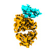
| Entry | Database: PDB / ID: 8crb | ||||||
|---|---|---|---|---|---|---|---|
| Title | Cryo-EM structure of PcrV/Fab(11-E5) | ||||||
 Components Components |
| ||||||
 Keywords Keywords |  ANTIMICROBIAL PROTEIN / antibody T3SS Tip protein ANTIMICROBIAL PROTEIN / antibody T3SS Tip protein | ||||||
| Function / homology |  Function and homology informationtype III protein secretion system complex / protein secretion by the type III secretion system / detection of maltose stimulus / maltose binding / maltose transport complex / maltose transport / maltodextrin transmembrane transport / carbohydrate transport / carbohydrate transmembrane transporter activity / ATP-binding cassette (ABC) transporter complex, substrate-binding subunit-containing ...type III protein secretion system complex / protein secretion by the type III secretion system / detection of maltose stimulus / maltose binding / maltose transport complex / maltose transport / maltodextrin transmembrane transport / carbohydrate transport / carbohydrate transmembrane transporter activity / ATP-binding cassette (ABC) transporter complex, substrate-binding subunit-containing / ATP-binding cassette (ABC) transporter complex / cell chemotaxis / outer membrane-bounded periplasmic space / periplasmic space / DNA damage response / extracellular space / membrane Function and homology informationtype III protein secretion system complex / protein secretion by the type III secretion system / detection of maltose stimulus / maltose binding / maltose transport complex / maltose transport / maltodextrin transmembrane transport / carbohydrate transport / carbohydrate transmembrane transporter activity / ATP-binding cassette (ABC) transporter complex, substrate-binding subunit-containing ...type III protein secretion system complex / protein secretion by the type III secretion system / detection of maltose stimulus / maltose binding / maltose transport complex / maltose transport / maltodextrin transmembrane transport / carbohydrate transport / carbohydrate transmembrane transporter activity / ATP-binding cassette (ABC) transporter complex, substrate-binding subunit-containing / ATP-binding cassette (ABC) transporter complex / cell chemotaxis / outer membrane-bounded periplasmic space / periplasmic space / DNA damage response / extracellular space / membraneSimilarity search - Function | ||||||
| Biological species |  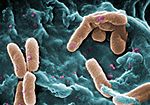 Pseudomonas aeruginosa (bacteria) Pseudomonas aeruginosa (bacteria) Homo sapiens (human) Homo sapiens (human) | ||||||
| Method | ELECTRON MICROSCOPY / single particle reconstruction / cryo EM / Resolution: 4.6 Å | ||||||
 Authors Authors | Yuan, B. / Simonis, A. / Marlovits, T.C. | ||||||
| Funding support |  Germany, 1items Germany, 1items
| ||||||
 Citation Citation | Journal: Cell / Year: 2023 Title: Discovery of highly neutralizing human antibodies targeting Pseudomonas aeruginosa. Authors: Alexander Simonis / Christoph Kreer / Alexandra Albus / Katharina Rox / Biao Yuan / Dmitriy Holzmann / Joana A Wilms / Sylvia Zuber / Lisa Kottege / Sandra Winter / Meike Meyer / Kristin ...Authors: Alexander Simonis / Christoph Kreer / Alexandra Albus / Katharina Rox / Biao Yuan / Dmitriy Holzmann / Joana A Wilms / Sylvia Zuber / Lisa Kottege / Sandra Winter / Meike Meyer / Kristin Schmitt / Henning Gruell / Sebastian J Theobald / Anna-Maria Hellmann / Christina Meyer / Meryem Seda Ercanoglu / Nina Cramer / Antje Munder / Michael Hallek / Gerd Fätkenheuer / Manuel Koch / Harald Seifert / Ernst Rietschel / Thomas C Marlovits / Silke van Koningsbruggen-Rietschel / Florian Klein / Jan Rybniker / Abstract: Drug-resistant Pseudomonas aeruginosa (PA) poses an emerging threat to human health with urgent need for alternative therapeutic approaches. Here, we deciphered the B cell and antibody response to ...Drug-resistant Pseudomonas aeruginosa (PA) poses an emerging threat to human health with urgent need for alternative therapeutic approaches. Here, we deciphered the B cell and antibody response to the virulence-associated type III secretion system (T3SS) in a cohort of patients chronically infected with PA. Single-cell analytics revealed a diverse B cell receptor repertoire directed against the T3SS needle-tip protein PcrV, enabling the production of monoclonal antibodies (mAbs) abrogating T3SS-mediated cytotoxicity. Mechanistic studies involving cryoelectron microscopy identified a surface-exposed C-terminal PcrV epitope as the target of highly neutralizing mAbs with broad activity against drug-resistant PA isolates. These anti-PcrV mAbs were as effective as treatment with conventional antibiotics in vivo. Our study reveals that chronically infected patients represent a source of neutralizing antibodies, which can be exploited as therapeutics against PA. | ||||||
| History |
|
- Structure visualization
| Structure viewer | Molecule: MolmilJmol/JSmol |
|---|
- Downloads & links
-Download
| PDBx/mmCIF format | 8crb.cif.gz | 225 KB | Display | PDBx/mmCIF format |
|---|---|---|---|---|
| PDB format | pdb8crb.ent.gz | 176.2 KB | Display | PDB format |
| PDBx/mmJSON format | 8crb.json.gz | Tree view | PDBx/mmJSON format | |
| Others |  Other downloads Other downloads |
-Validation report
| Arichive directory | https://data.pdbj.org/pub/pdb/validation_reports/cr/8crbftp://data.pdbj.org/pub/pdb/validation_reports/cr/8crb | HTTPS FTP |
|---|
-Related structure data
| Related structure data |  16807MC  8cr9C M: map data used to model this data C: citing same article ( |
|---|---|
| Similar structure data |
-Links
 PDBj
PDBj


- Assembly
| Deposited unit | 
|
|---|---|
| 1 |
|
-Components
| #1: Antibody | Mass: 24036.674 Da / Num. of mol.: 1 / Source method: isolated from a natural source / Source: (natural) Homo sapiens (human) |
|---|---|
| #2: Antibody | Mass: 22964.422 Da / Num. of mol.: 1 / Source method: isolated from a natural source / Source: (natural) Homo sapiens (human) |
| #3: Protein | Type three secretion system / MMBP / Maltodextrin-binding protein / Maltose-binding protein / MBP Mass: 75538.055 Da / Num. of mol.: 1 Source method: isolated from a genetically manipulated source Source: (gene. exp.) Pseudomonas aeruginosa (bacteria) / Gene: malE, b4034, JW3994, pcrV, PA1706 / Production host: Escherichia coli (E. coli) / References: UniProt: P0AEX9, UniProt: G3XD49 |
-Experimental details
-Experiment
| Experiment | Method: ELECTRON MICROSCOPY |
|---|---|
| EM experiment | Aggregation state: PARTICLE / 3D reconstruction method: single particle reconstruction |
- Sample preparation
| Component | Name: PcrV-Fab(11-E5) / Type: COMPLEX / Entity ID: all / Source: NATURAL |
|---|---|
| Molecular weight | Experimental value: NO |
| Source (natural) | Organism: Homo sapiens (human) |
| Source (recombinant) | Organism: Homo sapiens (human) |
| Buffer solution | pH: 7.4 / Details: 1xPBS |
| Specimen | Conc.: 0.33 mg/ml / Embedding applied: NO / Shadowing applied: NO / Staining applied: NO / Vitrification applied: YES |
| Vitrification | Cryogen name: ETHANE-PROPANE / Humidity: 100 % |
- Electron microscopy imaging
| Experimental equipment |  Model: Titan Krios / Image courtesy: FEI Company |
|---|---|
| Microscopy | Model: FEI TITAN KRIOS |
| Electron gun | Electron source: FIELD EMISSION GUN / Accelerating voltage: 300 kV / Illumination mode: FLOOD BEAM |
| Electron lens | Mode: BRIGHT FIELDBright-field microscopy / Nominal defocus max: 2500 nm / Nominal defocus min: 1000 nm |
| Image recording | Electron dose: 1.24 e/Å2 / Film or detector model: GATAN K3 (6k x 4k) |
- Processing
| Software | Name: UCSF ChimeraX / Version: 1.4/v9 / Classification: model building / URL: https://www.rbvi.ucsf.edu/chimerax/ / Os: Windows / Type: package |
|---|---|
| CTF correction | Type: PHASE FLIPPING AND AMPLITUDE CORRECTION |
| 3D reconstruction | Resolution: 4.6 Å / Resolution method: FSC 0.143 CUT-OFF / Num. of particles: 99625 / Symmetry type: POINT |
