 Movie
Movie Controller

[English] 日本語
 Yorodumi
- PDB-8ajl: Structure of the Ancestral Scaffold Antigen-6 of Coronavirus Spik... -
+ Open data

- Basic information
| Entry | Database: PDB / ID: 8ajl | ||||||||||||||||||
|---|---|---|---|---|---|---|---|---|---|---|---|---|---|---|---|---|---|---|---|
| Title | Structure of the Ancestral Scaffold Antigen-6 of Coronavirus Spike protein | ||||||||||||||||||
 Components Components | Spike glycoprotein,Fibritin | ||||||||||||||||||
 Keywords Keywords |  BIOSYNTHETIC PROTEIN / Ancestor / Spike / Coronavirus / Scaffold / S-protein / Protein Engineering BIOSYNTHETIC PROTEIN / Ancestor / Spike / Coronavirus / Scaffold / S-protein / Protein Engineering | ||||||||||||||||||
| Function / homology |  Function and homology informationvirion component / Maturation of spike protein / viral translation / Translation of Structural Proteins / Virion Assembly and Release / host cell surface / host extracellular space / suppression by virus of host tetherin activity / Induction of Cell-Cell Fusion / structural constituent of virion ...virion component / Maturation of spike protein / viral translation / Translation of Structural Proteins / Virion Assembly and Release / host cell surface / host extracellular space / suppression by virus of host tetherin activity / Induction of Cell-Cell Fusion / structural constituent of virion / entry receptor-mediated virion attachment to host cell / host cell endoplasmic reticulum-Golgi intermediate compartment membrane / receptor-mediated endocytosis of virus by host cell / Attachment and Entry / membrane fusion / positive regulation of viral entry into host cell / receptor-mediated virion attachment to host cell / receptor ligand activity / host cell surface receptor binding / fusion of virus membrane with host plasma membrane / fusion of virus membrane with host endosome membrane / viral envelope / symbiont-mediated suppression of host type I interferon-mediated signaling pathway / virion attachment to host cell / SARS-CoV-2 activates/modulates innate and adaptive immune responses / host cell plasma membrane / virion membrane / membrane / identical protein binding / plasma membrane Function and homology informationvirion component / Maturation of spike protein / viral translation / Translation of Structural Proteins / Virion Assembly and Release / host cell surface / host extracellular space / suppression by virus of host tetherin activity / Induction of Cell-Cell Fusion / structural constituent of virion ...virion component / Maturation of spike protein / viral translation / Translation of Structural Proteins / Virion Assembly and Release / host cell surface / host extracellular space / suppression by virus of host tetherin activity / Induction of Cell-Cell Fusion / structural constituent of virion / entry receptor-mediated virion attachment to host cell / host cell endoplasmic reticulum-Golgi intermediate compartment membrane / receptor-mediated endocytosis of virus by host cell / Attachment and Entry / membrane fusion / positive regulation of viral entry into host cell / receptor-mediated virion attachment to host cell / receptor ligand activity / host cell surface receptor binding / fusion of virus membrane with host plasma membrane / fusion of virus membrane with host endosome membrane / viral envelope / symbiont-mediated suppression of host type I interferon-mediated signaling pathway / virion attachment to host cell / SARS-CoV-2 activates/modulates innate and adaptive immune responses / host cell plasma membrane / virion membrane / membrane / identical protein binding / plasma membraneSimilarity search - Function | ||||||||||||||||||
| Biological species |  Severe acute respiratory syndrome coronavirusEnterobacteria phage T4 (virus) Severe acute respiratory syndrome coronavirusEnterobacteria phage T4 (virus) | ||||||||||||||||||
| Method | ELECTRON MICROSCOPY / single particle reconstruction / cryo EM / Resolution: 2.77 Å | ||||||||||||||||||
 Authors Authors | Hueting, D. / Schriever, K. / Wallden, K. / Andrell, J. / Syren, P.O. | ||||||||||||||||||
| Funding support |  Sweden, European Union, 5items Sweden, European Union, 5items
| ||||||||||||||||||
 Citation Citation | Journal: Nat Commun / Year: 2023 Title: Design, structure and plasma binding of ancestral β-CoV scaffold antigens. Authors: David Hueting / Karen Schriever / Rui Sun / Stelios Vlachiotis / Fanglei Zuo / Likun Du / Helena Persson / Camilla Hofström / Mats Ohlin / Karin Walldén / Marcus Buggert / Lennart ...Authors: David Hueting / Karen Schriever / Rui Sun / Stelios Vlachiotis / Fanglei Zuo / Likun Du / Helena Persson / Camilla Hofström / Mats Ohlin / Karin Walldén / Marcus Buggert / Lennart Hammarström / Harold Marcotte / Qiang Pan-Hammarström / Juni Andréll / Per-Olof Syrén / Abstract: We report the application of ancestral sequence reconstruction on coronavirus spike protein, resulting in stable and highly soluble ancestral scaffold antigens (AnSAs). The AnSAs interact with plasma ...We report the application of ancestral sequence reconstruction on coronavirus spike protein, resulting in stable and highly soluble ancestral scaffold antigens (AnSAs). The AnSAs interact with plasma of patients recovered from COVID-19 but do not bind to the human angiotensin-converting enzyme 2 (ACE2) receptor. Cryo-EM analysis of the AnSAs yield high resolution structures (2.6-2.8 Å) indicating a closed pre-fusion conformation in which all three receptor-binding domains (RBDs) are facing downwards. The structures reveal an intricate hydrogen-bonding network mediated by well-resolved loops, both within and across monomers, tethering the N-terminal domain and RBD together. We show that AnSA-5 can induce and boost a broad-spectrum immune response against the wild-type RBD as well as circulating variants of concern in an immune organoid model derived from tonsils. Finally, we highlight how AnSAs are potent scaffolds by replacing the ancestral RBD with the wild-type sequence, which restores ACE2 binding and increases the interaction with convalescent plasma. #1: Journal: Res Sq / Year: 2023Title: Design, structure and plasma binding of ancestral beta-CoV scaffold antigens Authors: Hueting, D. / Schriever, K. / Zuo, F. / Du, L. / Persson, H. / Hofstrom, C. / Ohlin, M. / Wallden, K. / Hammarstrom, L. / Marcotte, H. / Pan-Hammarstrom, Q. / Andrell, J. / Syren, P.O. | ||||||||||||||||||
| History |
|
- Structure visualization
| Structure viewer | Molecule: MolmilJmol/JSmol |
|---|
- Downloads & links
-Download
| PDBx/mmCIF format | 8ajl.cif.gz | 1.1 MB | Display | PDBx/mmCIF format |
|---|---|---|---|---|
| PDB format | pdb8ajl.ent.gz | Display | PDB format | |
| PDBx/mmJSON format | 8ajl.json.gz | Tree view | PDBx/mmJSON format | |
| Others |  Other downloads Other downloads |
-Validation report
| Arichive directory | https://data.pdbj.org/pub/pdb/validation_reports/aj/8ajlftp://data.pdbj.org/pub/pdb/validation_reports/aj/8ajl | HTTPS FTP |
|---|
-Related structure data
| Related structure data |  15482MC 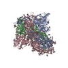 8ajaC C: citing same article ( M: map data used to model this data |
|---|---|
| Similar structure data |

-Links
 PDBj
PDBj


- Assembly
| Deposited unit | 
|
|---|---|
| 1 |
|
-Components
| #1: Protein | Mass: 138993.469 Da / Num. of mol.: 3 Source method: isolated from a genetically manipulated source Details: AnSA-6 full sequence 1-19 WT SARS-CoV-2 S protein signaling peptide 672-679 No density. 1139 onwards, No density. Expression tags.,AnSA-6 full sequence 1-19 WT SARS-CoV-2 S protein signaling ...Details: AnSA-6 full sequence 1-19 WT SARS-CoV-2 S protein signaling peptide 672-679 No density. 1139 onwards, No density. Expression tags.,AnSA-6 full sequence 1-19 WT SARS-CoV-2 S protein signaling peptide 672-679 No density. 1139 onwards, No density. Expression tags. Source: (gene. exp.) Severe acute respiratory syndrome coronavirus, (gene. exp.) Enterobacteria phage T4 (Bacteriophage T4) (virus)Gene: S, 2, wac / Plasmid: pMx series / Production host:  Homo sapiens (human) / Strain (production host): Expi293F / References: UniProt: P0DTC2, UniProt: P10104 Homo sapiens (human) / Strain (production host): Expi293F / References: UniProt: P0DTC2, UniProt: P10104#2: Polysaccharide | 2-acetamido-2-deoxy-beta-D-glucopyranose-(1-4)-2-acetamido-2-deoxy-beta-D-glucopyranose / Mass: 424.401 Da / Num. of mol.: 9Source method: isolated from a genetically manipulated source #3: Sugar | ChemComp-NAG / N-Acetylglucosamine  Type: D-saccharide, beta linking / Mass: 221.208 Da / Num. of mol.: 45 / Source method: obtained synthetically / Formula: C8H15NO6 / Feature type: SUBJECT OF INVESTIGATION Type: D-saccharide, beta linking / Mass: 221.208 Da / Num. of mol.: 45 / Source method: obtained synthetically / Formula: C8H15NO6 / Feature type: SUBJECT OF INVESTIGATIONHas ligand of interest | Y | |
|---|
-Experimental details
-Experiment
| Experiment | Method: ELECTRON MICROSCOPY |
|---|---|
| EM experiment | Aggregation state: PARTICLE / 3D reconstruction method: single particle reconstruction |
- Sample preparation
| Component | Name: Ancestral S-protein of coronaviruses related to SARS-CoV-2 Type: COMPLEX Details: Ancestral coronavirus generated by Ancestral sequence reconstruction expressed in human cells. Entity ID: #1 / Source: RECOMBINANT | |||||||||||||||
|---|---|---|---|---|---|---|---|---|---|---|---|---|---|---|---|---|
| Molecular weight | Experimental value: NO | |||||||||||||||
| Source (natural) |
| |||||||||||||||
| Source (recombinant) |
| |||||||||||||||
| Buffer solution | pH: 7.5 | |||||||||||||||
| Buffer component |
| |||||||||||||||
| Specimen | Conc.: 1.5 mg/ml / Embedding applied: NO / Shadowing applied: NO / Staining applied: NO / Vitrification applied: YES | |||||||||||||||
| Vitrification | Instrument: FEI VITROBOT MARK IV / Cryogen name: ETHANE / Humidity: 100 % / Chamber temperature: 277 K |
- Electron microscopy imaging
| Experimental equipment |  Model: Titan Krios / Image courtesy: FEI Company |
|---|---|
| Microscopy | Model: TFS KRIOS |
| Electron gun | Electron source: FIELD EMISSION GUN / Accelerating voltage: 300 kV / Illumination mode: OTHER |
| Electron lens | Mode: BRIGHT FIELDBright-field microscopy / Nominal magnification: 105000 X / Nominal defocus max: 2000 nm / Nominal defocus min: 600 nm / Cs: 2.7 mm / C2 aperture diameter: 50 µm |
| Specimen holder | Cryogen: NITROGEN / Specimen holder model: FEI TITAN KRIOS AUTOGRID HOLDER |
| Image recording | Electron dose: 1.11 e/Å2 / Film or detector model: GATAN K3 BIOQUANTUM (6k x 4k) |
| EM imaging optics | Energyfilter name: GIF Bioquantum / Energyfilter slit width: 20 eV |
- Processing
| Software | Name: PHENIX / Version: 1.19.2_4158: / Classification: refinement | ||||||||||||||||||||||||||||
|---|---|---|---|---|---|---|---|---|---|---|---|---|---|---|---|---|---|---|---|---|---|---|---|---|---|---|---|---|---|
| EM software |
| ||||||||||||||||||||||||||||
| CTF correction | Type: PHASE FLIPPING AND AMPLITUDE CORRECTION | ||||||||||||||||||||||||||||
| Particle selection | Num. of particles selected: 2035826 | ||||||||||||||||||||||||||||
| 3D reconstruction | Resolution: 2.77 Å / Resolution method: FSC 0.143 CUT-OFF / Num. of particles: 253429 / Symmetry type: POINT | ||||||||||||||||||||||||||||
| Atomic model building |
| ||||||||||||||||||||||||||||
| Refine LS restraints |
|


