[English] 日本語
 Yorodumi
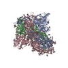
- EMDB-15475: Structure of the Ancestral Scaffold Antigen-5 of Coronavirus Spik... -
+ Open data
- Basic information
| Entry |  | ||||||||||||||||||
|---|---|---|---|---|---|---|---|---|---|---|---|---|---|---|---|---|---|---|---|
| Title | Structure of the Ancestral Scaffold Antigen-5 of Coronavirus Spike protein | ||||||||||||||||||
 Map data Map data | |||||||||||||||||||
 Sample Sample |
| ||||||||||||||||||
 Keywords Keywords |  Ancestor / Spike / Ancestor / Spike /  Coronavirus / Coronavirus /  Scaffold / S-protein / Scaffold / S-protein /  Protein Engineering / Protein Engineering /  BIOSYNTHETIC PROTEIN BIOSYNTHETIC PROTEIN | ||||||||||||||||||
| Function / homology |  Function and homology information Function and homology information virion component / Maturation of spike protein / viral translation / Translation of Structural Proteins / Virion Assembly and Release / host cell surface / host extracellular space / suppression by virus of host tetherin activity / Induction of Cell-Cell Fusion / structural constituent of virion ... virion component / Maturation of spike protein / viral translation / Translation of Structural Proteins / Virion Assembly and Release / host cell surface / host extracellular space / suppression by virus of host tetherin activity / Induction of Cell-Cell Fusion / structural constituent of virion ... virion component / Maturation of spike protein / viral translation / Translation of Structural Proteins / Virion Assembly and Release / host cell surface / host extracellular space / suppression by virus of host tetherin activity / Induction of Cell-Cell Fusion / structural constituent of virion / entry receptor-mediated virion attachment to host cell / host cell endoplasmic reticulum-Golgi intermediate compartment membrane / receptor-mediated endocytosis of virus by host cell / Attachment and Entry / virion component / Maturation of spike protein / viral translation / Translation of Structural Proteins / Virion Assembly and Release / host cell surface / host extracellular space / suppression by virus of host tetherin activity / Induction of Cell-Cell Fusion / structural constituent of virion / entry receptor-mediated virion attachment to host cell / host cell endoplasmic reticulum-Golgi intermediate compartment membrane / receptor-mediated endocytosis of virus by host cell / Attachment and Entry /  membrane fusion / positive regulation of viral entry into host cell / receptor-mediated virion attachment to host cell / membrane fusion / positive regulation of viral entry into host cell / receptor-mediated virion attachment to host cell /  receptor ligand activity / host cell surface receptor binding / fusion of virus membrane with host plasma membrane / fusion of virus membrane with host endosome membrane / receptor ligand activity / host cell surface receptor binding / fusion of virus membrane with host plasma membrane / fusion of virus membrane with host endosome membrane /  viral envelope / symbiont-mediated suppression of host type I interferon-mediated signaling pathway / virion attachment to host cell / SARS-CoV-2 activates/modulates innate and adaptive immune responses / host cell plasma membrane / virion membrane / viral envelope / symbiont-mediated suppression of host type I interferon-mediated signaling pathway / virion attachment to host cell / SARS-CoV-2 activates/modulates innate and adaptive immune responses / host cell plasma membrane / virion membrane /  membrane / identical protein binding / membrane / identical protein binding /  plasma membrane plasma membraneSimilarity search - Function | ||||||||||||||||||
| Biological species |   Severe acute respiratory syndrome coronavirus / Severe acute respiratory syndrome coronavirus /   Tequatrovirus T4 Tequatrovirus T4 | ||||||||||||||||||
| Method |  single particle reconstruction / single particle reconstruction /  cryo EM / Resolution: 2.59 Å cryo EM / Resolution: 2.59 Å | ||||||||||||||||||
 Authors Authors | Hueting D / Schriever K / Wallden K / Andrell J / Syren PO | ||||||||||||||||||
| Funding support |  Sweden, European Union, 5 items Sweden, European Union, 5 items
| ||||||||||||||||||
 Citation Citation |  Journal: Nat Commun / Year: 2023 Journal: Nat Commun / Year: 2023Title: Design, structure and plasma binding of ancestral β-CoV scaffold antigens. Authors: David Hueting / Karen Schriever / Rui Sun / Stelios Vlachiotis / Fanglei Zuo / Likun Du / Helena Persson / Camilla Hofström / Mats Ohlin / Karin Walldén / Marcus Buggert / Lennart ...Authors: David Hueting / Karen Schriever / Rui Sun / Stelios Vlachiotis / Fanglei Zuo / Likun Du / Helena Persson / Camilla Hofström / Mats Ohlin / Karin Walldén / Marcus Buggert / Lennart Hammarström / Harold Marcotte / Qiang Pan-Hammarström / Juni Andréll / Per-Olof Syrén /  Abstract: We report the application of ancestral sequence reconstruction on coronavirus spike protein, resulting in stable and highly soluble ancestral scaffold antigens (AnSAs). The AnSAs interact with plasma ...We report the application of ancestral sequence reconstruction on coronavirus spike protein, resulting in stable and highly soluble ancestral scaffold antigens (AnSAs). The AnSAs interact with plasma of patients recovered from COVID-19 but do not bind to the human angiotensin-converting enzyme 2 (ACE2) receptor. Cryo-EM analysis of the AnSAs yield high resolution structures (2.6-2.8 Å) indicating a closed pre-fusion conformation in which all three receptor-binding domains (RBDs) are facing downwards. The structures reveal an intricate hydrogen-bonding network mediated by well-resolved loops, both within and across monomers, tethering the N-terminal domain and RBD together. We show that AnSA-5 can induce and boost a broad-spectrum immune response against the wild-type RBD as well as circulating variants of concern in an immune organoid model derived from tonsils. Finally, we highlight how AnSAs are potent scaffolds by replacing the ancestral RBD with the wild-type sequence, which restores ACE2 binding and increases the interaction with convalescent plasma. #1:  Journal: Res Sq / Year: 2023 Journal: Res Sq / Year: 2023Title: Design, structure and plasma binding of ancestral beta-CoV scaffold antigens Authors: Hueting D / Schriever K / Zuo F / Du L / Persson H / Hofstrom C / Ohlin M / Wallden K / Hammarstrom L / Marcotte H / Pan-Hammarstrom Q / Andrell J / Syren PO | ||||||||||||||||||
| History |
|
- Structure visualization
| Supplemental images |
|---|
- Downloads & links
-EMDB archive
| Map data |  emd_15475.map.gz emd_15475.map.gz | 204.1 MB |  EMDB map data format EMDB map data format | |
|---|---|---|---|---|
| Header (meta data) |  emd-15475-v30.xml emd-15475-v30.xml emd-15475.xml emd-15475.xml | 21.9 KB 21.9 KB | Display Display |  EMDB header EMDB header |
| FSC (resolution estimation) |  emd_15475_fsc.xml emd_15475_fsc.xml | 12.6 KB | Display |  FSC data file FSC data file |
| Images |  emd_15475.png emd_15475.png | 26.6 KB | ||
| Masks |  emd_15475_msk_1.map emd_15475_msk_1.map | 216 MB |  Mask map Mask map | |
| Filedesc metadata |  emd-15475.cif.gz emd-15475.cif.gz | 7.5 KB | ||
| Others |  emd_15475_half_map_1.map.gz emd_15475_half_map_1.map.gz emd_15475_half_map_2.map.gz emd_15475_half_map_2.map.gz | 200.8 MB 200.8 MB | ||
| Archive directory |  http://ftp.pdbj.org/pub/emdb/structures/EMD-15475 http://ftp.pdbj.org/pub/emdb/structures/EMD-15475 ftp://ftp.pdbj.org/pub/emdb/structures/EMD-15475 ftp://ftp.pdbj.org/pub/emdb/structures/EMD-15475 | HTTPS FTP |
-Related structure data
| Related structure data |  8ajaMC  8ajlC M: atomic model generated by this map C: citing same article ( |
|---|---|
| Similar structure data | Similarity search - Function & homology  F&H Search F&H Search |
- Links
Links
| EMDB pages |  EMDB (EBI/PDBe) / EMDB (EBI/PDBe) /  EMDataResource EMDataResource |
|---|---|
| Related items in Molecule of the Month |
- Map
Map
| File |  Download / File: emd_15475.map.gz / Format: CCP4 / Size: 216 MB / Type: IMAGE STORED AS FLOATING POINT NUMBER (4 BYTES) Download / File: emd_15475.map.gz / Format: CCP4 / Size: 216 MB / Type: IMAGE STORED AS FLOATING POINT NUMBER (4 BYTES) | ||||||||||||||||||||||||||||||||||||
|---|---|---|---|---|---|---|---|---|---|---|---|---|---|---|---|---|---|---|---|---|---|---|---|---|---|---|---|---|---|---|---|---|---|---|---|---|---|
| Projections & slices | Image control
Images are generated by Spider. | ||||||||||||||||||||||||||||||||||||
| Voxel size | X=Y=Z: 1.11 Å | ||||||||||||||||||||||||||||||||||||
| Density |
| ||||||||||||||||||||||||||||||||||||
| Symmetry | Space group: 1 | ||||||||||||||||||||||||||||||||||||
| Details | EMDB XML:
|
-Supplemental data
-Mask #1
| File |  emd_15475_msk_1.map emd_15475_msk_1.map | ||||||||||||
|---|---|---|---|---|---|---|---|---|---|---|---|---|---|
| Projections & Slices |
| ||||||||||||
| Density Histograms |
-Half map: #1
| File | emd_15475_half_map_1.map | ||||||||||||
|---|---|---|---|---|---|---|---|---|---|---|---|---|---|
| Projections & Slices |
| ||||||||||||
| Density Histograms |
-Half map: #2
| File | emd_15475_half_map_2.map | ||||||||||||
|---|---|---|---|---|---|---|---|---|---|---|---|---|---|
| Projections & Slices |
| ||||||||||||
| Density Histograms |
- Sample components
-Entire : Ancestral S-protein of coronaviruses related to SARS-CoV-2
| Entire | Name: Ancestral S-protein of coronaviruses related to SARS-CoV-2 |
|---|---|
| Components |
|
-Supramolecule #1: Ancestral S-protein of coronaviruses related to SARS-CoV-2
| Supramolecule | Name: Ancestral S-protein of coronaviruses related to SARS-CoV-2 type: complex / ID: 1 / Parent: 0 / Macromolecule list: #1 Details: Ancestral coronavirus generated by Ancestral sequence reconstruction expressed in human cells. |
|---|---|
| Source (natural) | Organism:   Severe acute respiratory syndrome coronavirus Severe acute respiratory syndrome coronavirus |
-Macromolecule #1: Spike glycoprotein,Fibritin
| Macromolecule | Name: Spike glycoprotein,Fibritin / type: protein_or_peptide / ID: 1 Details: AnSA-5 With His-Tag and Twin-Strep-tag and SARS-CoV-2 signaling peptide 1-19 Signaling peptide 653-660 No density 1119 onwards, no density and expression and purification tag,AnSA-5 With His- ...Details: AnSA-5 With His-Tag and Twin-Strep-tag and SARS-CoV-2 signaling peptide 1-19 Signaling peptide 653-660 No density 1119 onwards, no density and expression and purification tag,AnSA-5 With His-Tag and Twin-Strep-tag and SARS-CoV-2 signaling peptide 1-19 Signaling peptide 653-660 No density 1119 onwards, no density and expression and purification tag,AnSA-5 With His-Tag and Twin-Strep-tag and SARS-CoV-2 signaling peptide 1-19 Signaling peptide 653-660 No density 1119 onwards, no density and expression and purification tag,AnSA-5 With His-Tag and Twin-Strep-tag and SARS-CoV-2 signaling peptide 1-19 Signaling peptide 653-660 No density 1119 onwards, no density and expression and purification tag Number of copies: 3 / Enantiomer: LEVO |
|---|---|
| Source (natural) | Organism:   Tequatrovirus T4 Tequatrovirus T4 |
| Molecular weight | Theoretical: 137.212609 KDa |
| Recombinant expression | Organism:   Homo sapiens (human) Homo sapiens (human) |
| Sequence | String: TCGTISNKTP PNMNQFSSSR RGVYYPDDIF RSDVLHLTQD YFLPFNSNVT RYLSLNADSN RIVRFDNPIL PFGDGIYFAA TEKSNVIRG WIFGSTLDNT SQSAIIVNNS THIIIKVCNF QLCDDPMFTV SRGQHYKTWV YTNARNCTYE YVSKSFQLDV S EKNGNFKH ...String: TCGTISNKTP PNMNQFSSSR RGVYYPDDIF RSDVLHLTQD YFLPFNSNVT RYLSLNADSN RIVRFDNPIL PFGDGIYFAA TEKSNVIRG WIFGSTLDNT SQSAIIVNNS THIIIKVCNF QLCDDPMFTV SRGQHYKTWV YTNARNCTYE YVSKSFQLDV S EKNGNFKH LREFVFKNVD GFLHVYSAYE PIDLARGLPS GFSVLKPILK LPLGINITSF RVVMTMFSPT TSNWLAESAA YF VGYLKPT TFMLKFNENG TITDAVDCSQ DPLSELKCTL KSFNVEKGIY QTSNFRVSPT QEVVRFPNIT NLCPFDKVFN ATR FPSVYA WERTKISDCV ADYTVLYNST SFSTFKCYGV SPSKLIDLCF TSVYADTFLI RSSEVRQVAP GQTGVIADYN YKLP DDFTG CVIAWNTAKQ DAGNYYYRSH RKTKLKPFER DLSNSDENGV RTLSTYDFNP NVPIEYQATR VVVLSFELLN APATV CGPK LSTQLVKNQC VNFNFNGLKG TGVLTDSSKR FQSFQQFGRD ASDFTDSVRD PQTLEILDIS PCSFGGVSVI TPGTNA SSE VAVLYQDVNC TDVPTAIHAD QLTPAWRVYS TGVNVFQTQA GCLIGAEHVN ASYECDIPIG AGICASYHTA STLRSTG QK SIVAYTMSLG AENSIAYANN SIAIPTNFSI SVTTEVMPVS MAKTSVDCTM YICGDSQECS NLLLQYGSFC TQLNRALT G IAIEQDKNTQ EVFAQVKQMY KTPAIKDFGG FNFSQILPDP SKPTKRSFIE DLLFNKVTLA DAGFMKQYGE CLGDISARD LICAQKFNGL TVLPPLLTDE MIAAYTAALV SGTATAGWTF GAGAALQIPF AMQMAYRFNG IGVTQNVLYE NQKQIANQFN KAISQIQES LTTTSTALGK LQDVVNQNAQ ALNTLVKQLS SNFGAISSVL NDILSRLDKV EAEVQIDRLI TGRLQSLQTY V TQQLIRAA EIRASANLAA TKMSECVLGQ SKRVDFCGKG YHLMSFPQAA PHGVVFLHVT YVPSQERNFT TAPAICHEGK AY FPREGVF VSNGTSWFIT QRNFYSPQII TTDNTFVAGN CDVVIGIINN TVYDPLQPEL DSFKEELDKY FKNHTSPDVD LGD ISGINA SVVNIQKEID RLNEVAKNLN ESLIDLQELG KYEQGSGYIP EAPRDGQAYV RKDGEWVLLS TFLGTSLEVL FQGP GHHHH HHHHSAWSHP QFEKGGGSGG GGSGGSAWSH PQFEK UniProtKB:  Spike glycoprotein, Fibritin Spike glycoprotein, Fibritin |
-Macromolecule #3: 2-acetamido-2-deoxy-beta-D-glucopyranose
| Macromolecule | Name: 2-acetamido-2-deoxy-beta-D-glucopyranose / type: ligand / ID: 3 / Number of copies: 39 / Formula: NAG |
|---|---|
| Molecular weight | Theoretical: 221.208 Da |
| Chemical component information |  ChemComp-NAG: |
-Experimental details
-Structure determination
| Method |  cryo EM cryo EM |
|---|---|
 Processing Processing |  single particle reconstruction single particle reconstruction |
| Aggregation state | particle |
- Sample preparation
| Concentration | 1.5 mg/mL | |||||||||
|---|---|---|---|---|---|---|---|---|---|---|
| Buffer | pH: 7.5 Component:
| |||||||||
| Grid | Model: UltrAuFoil R0.6/1 / Material: GOLD / Mesh: 300 / Pretreatment - Type: GLOW DISCHARGE / Pretreatment - Time: 60 sec. / Pretreatment - Atmosphere: AIR Details: Grids were glow discharged with 20mA for 60s in GlowQube system. | |||||||||
| Vitrification | Cryogen name: ETHANE / Chamber humidity: 100 % / Chamber temperature: 277 K / Instrument: FEI VITROBOT MARK IV |
- Electron microscopy
| Microscope | TFS KRIOS |
|---|---|
| Electron beam | Acceleration voltage: 300 kV / Electron source:  FIELD EMISSION GUN FIELD EMISSION GUN |
| Electron optics | C2 aperture diameter: 50.0 µm / Illumination mode: OTHER / Imaging mode: BRIGHT FIELD Bright-field microscopy / Cs: 2.7 mm / Nominal defocus max: 2.0 µm / Nominal defocus min: 0.6 µm / Nominal magnification: 105000 Bright-field microscopy / Cs: 2.7 mm / Nominal defocus max: 2.0 µm / Nominal defocus min: 0.6 µm / Nominal magnification: 105000 |
| Specialist optics | Energy filter - Name: GIF Bioquantum / Energy filter - Slit width: 20 eV |
| Sample stage | Specimen holder model: FEI TITAN KRIOS AUTOGRID HOLDER / Cooling holder cryogen: NITROGEN |
| Image recording | Film or detector model: GATAN K3 BIOQUANTUM (6k x 4k) / Average electron dose: 1.11 e/Å2 |
| Experimental equipment |  Model: Titan Krios / Image courtesy: FEI Company |
 Movie
Movie Controller


 Z (Sec.)
 Y (Row.)
 X (Col.)


