 Movie
Movie Controller

[English] 日本語
 Yorodumi
- PDB-7s0h: Crystal structure of Penicillium verruculosum copalyl diphosphate... -
+ Open data

- Basic information
| Entry | Database: PDB / ID: 7s0h | |||||||||
|---|---|---|---|---|---|---|---|---|---|---|
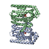
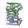
| Title | Crystal structure of Penicillium verruculosum copalyl diphosphate synthase (PvCPS) alpha prenyltransferase domain variant, S723Y | |||||||||
 Components Components | Terpene synthase | |||||||||
 Keywords Keywords | TRANSFERASE / Prenyltransferase / Isoprenoid Synthase / GGPP synthase / Bifunctional Terpene Synthase / Assembly-line synthase | |||||||||
| Function / homology |  Function and homology informationcopalyl diphosphate synthase / alcohol biosynthetic process / mycotoxin biosynthetic process / geranylgeranyl diphosphate synthase / isoprenoid biosynthetic process / isomerase activity / transferase activity / metal ion binding Function and homology informationcopalyl diphosphate synthase / alcohol biosynthetic process / mycotoxin biosynthetic process / geranylgeranyl diphosphate synthase / isoprenoid biosynthetic process / isomerase activity / transferase activity / metal ion bindingSimilarity search - Function | |||||||||
| Biological species |  Talaromyces verruculosus (fungus) Talaromyces verruculosus (fungus) | |||||||||
| Method | X-RAY DIFFRACTION / SYNCHROTRON / MOLECULAR REPLACEMENT / Resolution: 3.15 Å | |||||||||
 Authors Authors | Ronnebaum, T.A. / Christianson, D.W. | |||||||||
| Funding support |  United States, 2items United States, 2items
| |||||||||
 Citation Citation | Journal: Biochemistry / Year: 2021 Title: Engineering the Prenyltransferase Domain of a Bifunctional Assembly-Line Terpene Synthase. Authors: Ronnebaum, T.A. / Eaton, S.A. / Brackhahn, E.A.E. / Christianson, D.W. | |||||||||
| History |
|
- Structure visualization
| Structure viewer | Molecule: MolmilJmol/JSmol |
|---|
- Downloads & links
-Download
| PDBx/mmCIF format | 7s0h.cif.gz | 165.8 KB | Display | PDBx/mmCIF format |
|---|---|---|---|---|
| PDB format | pdb7s0h.ent.gz | 106.5 KB | Display | PDB format |
| PDBx/mmJSON format | 7s0h.json.gz | Tree view | PDBx/mmJSON format | |
| Others |  Other downloads Other downloads |
-Validation report
| Arichive directory | https://data.pdbj.org/pub/pdb/validation_reports/s0/7s0hftp://data.pdbj.org/pub/pdb/validation_reports/s0/7s0h | HTTPS FTP |
|---|
-Related structure data
| Related structure data | 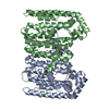 7s09C 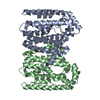 7s0aC 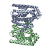 7s0lC  7s0mC  6v0kS S: Starting model for refinement C: citing same article ( |
|---|---|
| Similar structure data |


-Links
 PDBj
PDBj

- Assembly
| Deposited unit | 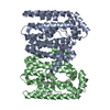
| ||||||||||||
|---|---|---|---|---|---|---|---|---|---|---|---|---|---|
| 1 | 
| ||||||||||||
| Unit cell |
|
-Components
| #1: Protein | Mass: 34979.109 Da / Num. of mol.: 2 / Fragment: Prenyltransferase alpha domain, residues 659-963 / Mutation: S723Y Source method: isolated from a genetically manipulated source Source: (gene. exp.) Talaromyces verruculosus (fungus) / Gene: PvCPS / Production host:  Escherichia coli BL21(DE3) (bacteria) / Strain (production host): BL21(DE3) / References: UniProt: A0A348FUE1 Escherichia coli BL21(DE3) (bacteria) / Strain (production host): BL21(DE3) / References: UniProt: A0A348FUE1Sequence details | The authors state that the full-length enzyme is a chimera, consisting of 963 residues. However, ...The authors state that the full-length enzyme is a chimera, consisting of 963 residues. However, the provided crystal structure was generated from limited proteolysis experiments and only the prenyltransferase alpha domain of the chimera was crystallized. The sequence of the full-length enzyme is: MSPMDLQESA | |
|---|
-Experimental details
-Experiment
| Experiment | Method: X-RAY DIFFRACTION / Number of used crystals: 1 |
|---|
- Sample preparation
| Crystal | Density Matthews: 4.14 Å3/Da / Density % sol: 70.28 % |
|---|---|
| Crystal grow | Temperature: 294 K / Method: vapor diffusion, hanging drop Details: 0.1 M sodium citrate pH 5.6 2.5% tacsimate pH 5.0 20% PEG 3350 |
-Data collection
| Diffraction | Mean temperature: 100 K / Serial crystal experiment: N |
|---|---|
| Diffraction source | Source: SYNCHROTRON / Site: APS / Beamline: 24-ID-E / Wavelength: 0.98 Å |
| Detector | Type: DECTRIS EIGER X 16M / Detector: PIXEL / Date: Jul 11, 2021 |
| Radiation | Protocol: SINGLE WAVELENGTH / Monochromatic (M) / Laue (L): M / Scattering type: x-ray |
| Radiation wavelength | Wavelength: 0.98 Å / Relative weight: 1 |
| Reflection | Resolution: 3.15→81.64 Å / Num. obs: 143593 / % possible obs: 100 % / Redundancy: 7.1 % / Biso Wilson estimate: 37.62 Å2 / CC1/2: 0.947 / Net I/σ(I): 5.3 |
| Reflection shell | Resolution: 3.15→3.263 Å / Mean I/σ(I) obs: 2 / Num. unique obs: 20131 / CC1/2: 0.509 |
- Processing
| Software |
| |||||||||||||||||||||||||||||||||||||||||||||||||||||||||||||||||||||||||||||||||||||||||||||||||||||||||||||||||||||||||||||||||||||||||||||||||||||||||||||||||||||||||||||||||||||||||||||||||||||||||||
|---|---|---|---|---|---|---|---|---|---|---|---|---|---|---|---|---|---|---|---|---|---|---|---|---|---|---|---|---|---|---|---|---|---|---|---|---|---|---|---|---|---|---|---|---|---|---|---|---|---|---|---|---|---|---|---|---|---|---|---|---|---|---|---|---|---|---|---|---|---|---|---|---|---|---|---|---|---|---|---|---|---|---|---|---|---|---|---|---|---|---|---|---|---|---|---|---|---|---|---|---|---|---|---|---|---|---|---|---|---|---|---|---|---|---|---|---|---|---|---|---|---|---|---|---|---|---|---|---|---|---|---|---|---|---|---|---|---|---|---|---|---|---|---|---|---|---|---|---|---|---|---|---|---|---|---|---|---|---|---|---|---|---|---|---|---|---|---|---|---|---|---|---|---|---|---|---|---|---|---|---|---|---|---|---|---|---|---|---|---|---|---|---|---|---|---|---|---|---|---|---|---|---|---|---|
| Refinement | Method to determine structure: MOLECULAR REPLACEMENT Starting model: 6V0K Resolution: 3.15→81.64 Å / SU ML: 0.407 / Cross valid method: FREE R-VALUE / σ(F): 1.34 / Phase error: 21.9922 Stereochemistry target values: GeoStd + Monomer Library + CDL v1.2
| |||||||||||||||||||||||||||||||||||||||||||||||||||||||||||||||||||||||||||||||||||||||||||||||||||||||||||||||||||||||||||||||||||||||||||||||||||||||||||||||||||||||||||||||||||||||||||||||||||||||||||
| Solvent computation | Shrinkage radii: 0.9 Å / VDW probe radii: 1.11 Å / Solvent model: FLAT BULK SOLVENT MODEL | |||||||||||||||||||||||||||||||||||||||||||||||||||||||||||||||||||||||||||||||||||||||||||||||||||||||||||||||||||||||||||||||||||||||||||||||||||||||||||||||||||||||||||||||||||||||||||||||||||||||||||
| Displacement parameters | Biso mean: 29.41 Å2 | |||||||||||||||||||||||||||||||||||||||||||||||||||||||||||||||||||||||||||||||||||||||||||||||||||||||||||||||||||||||||||||||||||||||||||||||||||||||||||||||||||||||||||||||||||||||||||||||||||||||||||
| Refinement step | Cycle: LAST / Resolution: 3.15→81.64 Å
| |||||||||||||||||||||||||||||||||||||||||||||||||||||||||||||||||||||||||||||||||||||||||||||||||||||||||||||||||||||||||||||||||||||||||||||||||||||||||||||||||||||||||||||||||||||||||||||||||||||||||||
| Refine LS restraints |
| |||||||||||||||||||||||||||||||||||||||||||||||||||||||||||||||||||||||||||||||||||||||||||||||||||||||||||||||||||||||||||||||||||||||||||||||||||||||||||||||||||||||||||||||||||||||||||||||||||||||||||
| LS refinement shell |
| |||||||||||||||||||||||||||||||||||||||||||||||||||||||||||||||||||||||||||||||||||||||||||||||||||||||||||||||||||||||||||||||||||||||||||||||||||||||||||||||||||||||||||||||||||||||||||||||||||||||||||
| Refinement TLS params. | Method: refined / Refine-ID: X-RAY DIFFRACTION
| |||||||||||||||||||||||||||||||||||||||||||||||||||||||||||||||||||||||||||||||||||||||||||||||||||||||||||||||||||||||||||||||||||||||||||||||||||||||||||||||||||||||||||||||||||||||||||||||||||||||||||
| Refinement TLS group | Refine-ID: X-RAY DIFFRACTION / Auth seq-ID: 858 - 870
|
