 Movie
Movie Controller

[English] 日本語
 Yorodumi
- PDB-7pg1: Crystal Structure of Unlinked NS2B-NS3 Protease from Zika Virus i... -
+ Open data

- Basic information
| Entry | Database: PDB / ID: 7pg1 | ||||||
|---|---|---|---|---|---|---|---|
| Title | Crystal Structure of Unlinked NS2B-NS3 Protease from Zika Virus in Complex with Inhibitor MI-2221 | ||||||
 Components Components |
| ||||||
 Keywords Keywords |  VIRAL PROTEIN / FLAVIVIRIN / SERINE PROTEASE / NS2B-NS3 / ZIKA VIRUS VIRAL PROTEIN / FLAVIVIRIN / SERINE PROTEASE / NS2B-NS3 / ZIKA VIRUS | ||||||
| Function / homology |  Function and homology information Function and homology informationsymbiont-mediated suppression of host JAK-STAT cascade via inhibition of host TYK2 activity / flavivirin / symbiont-mediated suppression of host JAK-STAT cascade via inhibition of STAT2 activity / symbiont-mediated suppression of host JAK-STAT cascade via inhibition of STAT1 activity / viral capsid / nucleoside-triphosphate phosphatase / double-stranded RNA binding / symbiont-mediated suppression of host cytoplasmic pattern recognition receptor signaling pathway via inhibition of TBK1 activity / symbiont-mediated suppression of host toll-like receptor signaling pathway / mRNA (guanine-N7)-methyltransferase ...symbiont-mediated suppression of host JAK-STAT cascade via inhibition of host TYK2 activity / flavivirin / symbiont-mediated suppression of host JAK-STAT cascade via inhibition of STAT2 activity / symbiont-mediated suppression of host JAK-STAT cascade via inhibition of STAT1 activity / viral capsid / nucleoside-triphosphate phosphatase / double-stranded RNA binding / symbiont-mediated suppression of host cytoplasmic pattern recognition receptor signaling pathway via inhibition of TBK1 activity / symbiont-mediated suppression of host toll-like receptor signaling pathway / mRNA (guanine-N7)-methyltransferase / methyltransferase cap1 / clathrin-dependent endocytosis of virus by host cell / mRNA (nucleoside-2'-O-)-methyltransferase activity / mRNA 5'-cap (guanine-N7-)-methyltransferase activity / RNA helicase activity / host cell endoplasmic reticulum membrane / host cell perinuclear region of cytoplasm / protein dimerization activity / RNA helicase / induction by virus of host autophagy / RNA-directed RNA polymerase / viral RNA genome replication / RNA-dependent RNA polymerase activity / serine-type endopeptidase activity / fusion of virus membrane with host endosome membrane / viral envelope / lipid binding / symbiont-mediated suppression of host type I interferon-mediated signaling pathway / host cell nucleus / virion attachment to host cell / GTP binding / virion membrane / structural molecule activity / ATP hydrolysis activity / proteolysis / extracellular region / ATP binding / membrane / metal ion bindingSimilarity search - Function | ||||||
| Biological species |  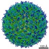 Zika virus Zika virussynhtetic construct (others) | ||||||
| Method | X-RAY DIFFRACTION / SYNCHROTRON / MOLECULAR REPLACEMENT / Resolution: 1.95 Å | ||||||
 Authors Authors | Huber, S. / Heine, A. / Steinmetzer, T. | ||||||
| Funding support | 1items
| ||||||
 Citation Citation | Journal: J.Med.Chem. / Year: 2022 Title: Structure-Based Optimization and Characterization of Macrocyclic Zika Virus NS2B-NS3 Protease Inhibitors. Authors: Huber, S. / Braun, N.J. / Schmacke, L.C. / Quek, J.P. / Murra, R. / Bender, D. / Hildt, E. / Luo, D. / Heine, A. / Steinmetzer, T. | ||||||
| History |
|
- Structure visualization
| Structure viewer | Molecule: MolmilJmol/JSmol |
|---|
- Downloads & links
-Download
| PDBx/mmCIF format | 7pg1.cif.gz | 99.9 KB | Display | PDBx/mmCIF format |
|---|---|---|---|---|
| PDB format | pdb7pg1.ent.gz | 61.3 KB | Display | PDB format |
| PDBx/mmJSON format | 7pg1.json.gz | Tree view | PDBx/mmJSON format | |
| Others |  Other downloads Other downloads |
-Validation report
| Arichive directory | https://data.pdbj.org/pub/pdb/validation_reports/pg/7pg1ftp://data.pdbj.org/pub/pdb/validation_reports/pg/7pg1 | HTTPS FTP |
|---|
-Related structure data
| Related structure data |  7o2mC 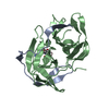 7o55C 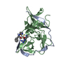 7obvC 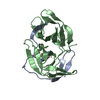 7oc2C  7pfqC  7pfyC  7pfzC  7pgcC 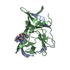 7vlgC 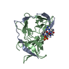 7vlhC  7vliC 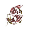 5gpiS S: Starting model for refinement C: citing same article ( |
|---|---|
| Similar structure data |
-Links
 PDBj
PDBj


- Assembly
| Deposited unit | 
| ||||||||||||
|---|---|---|---|---|---|---|---|---|---|---|---|---|---|
| 1 |
| ||||||||||||
| Unit cell |
| ||||||||||||
| Components on special symmetry positions |
|
-Components
| #1: Protein | / Flavivirin protease NS2B regulatory subunit / Non-structural protein 2B Mass: 5865.384 Da / Num. of mol.: 1 Source method: isolated from a genetically manipulated source Source: (gene. exp.) Zika virus / Plasmid: bZiPro / Production host:  Escherichia coli BL21(DE3) (bacteria) / References: UniProt: Q32ZE1 Escherichia coli BL21(DE3) (bacteria) / References: UniProt: Q32ZE1 |
|---|---|
| #2: Protein | Mass: 19037.592 Da / Num. of mol.: 1 Source method: isolated from a genetically manipulated source Source: (gene. exp.) Zika virus / Plasmid: bZiPro / Production host: Escherichia coli BL21(DE3) (bacteria)References: UniProt: Q32ZE1, flavivirin, nucleoside-triphosphate phosphatase, RNA helicase |
| #3: Protein/peptide | Mass: 631.769 Da / Num. of mol.: 1 / Source method: obtained synthetically / Source: (synth.) synhtetic construct (others) |
| #4: Water | ChemComp-HOH / Water Mass: 18.015 Da / Num. of mol.: 68 / Source method: isolated from a natural source / Formula: H2O Mass: 18.015 Da / Num. of mol.: 68 / Source method: isolated from a natural source / Formula: H2O |
| Has ligand of interest | Y |
-Experimental details
-Experiment
| Experiment | Method: X-RAY DIFFRACTION / Number of used crystals: 1 |
|---|
- Sample preparation
| Crystal | Density Matthews: 1.96 Å3/Da / Density % sol: 37.37 % |
|---|---|
| Crystal grow | Temperature: 291 K / Method: vapor diffusion, hanging drop / pH: 4.6 Details: 0.1 M sodium acetate 0.2 M ammonium sulfate 17% PEG2000 |
-Data collection
| Diffraction | Mean temperature: 100 K / Serial crystal experiment: N |
|---|---|
| Diffraction source | Source: SYNCHROTRON / Site: BESSY  / Beamline: 14.2 / Wavelength: 0.9184 Å / Beamline: 14.2 / Wavelength: 0.9184 Å |
| Detector | Type: DECTRIS PILATUS3 2M / Detector: PIXEL / Date: Nov 30, 2020 |
| Radiation | Protocol: SINGLE WAVELENGTH / Monochromatic (M) / Laue (L): M / Scattering type: x-ray |
| Radiation wavelength | Wavelength: 0.9184 Å / Relative weight: 1 |
| Reflection | Resolution: 1.95→43.1 Å / Num. obs: 15399 / % possible obs: 100 % / Redundancy: 14.29 % / Biso Wilson estimate: 35.55 Å2 / CC1/2: 1 / Rsym value: 0.053 / Net I/σ(I): 29.03 |
| Reflection shell | Resolution: 1.95→2.07 Å / Redundancy: 13.6 % / Mean I/σ(I) obs: 4.84 / Num. unique obs: 2415 / CC1/2: 0.982 / Rsym value: 0.517 / % possible all: 100 |
- Processing
| Software |
| ||||||||||||||||||||||||||||||||||||||||||
|---|---|---|---|---|---|---|---|---|---|---|---|---|---|---|---|---|---|---|---|---|---|---|---|---|---|---|---|---|---|---|---|---|---|---|---|---|---|---|---|---|---|---|---|
| Refinement | Method to determine structure: MOLECULAR REPLACEMENT Starting model: 5GPI Resolution: 1.95→36.59 Å / SU ML: 0.1966 / Cross valid method: FREE R-VALUE / σ(F): 1.37 / Phase error: 25.1514 Stereochemistry target values: GeoStd + Monomer Library + CDL v1.2
| ||||||||||||||||||||||||||||||||||||||||||
| Solvent computation | Shrinkage radii: 0.9 Å / VDW probe radii: 1.11 Å / Solvent model: FLAT BULK SOLVENT MODEL | ||||||||||||||||||||||||||||||||||||||||||
| Displacement parameters | Biso mean: 42.58 Å2 | ||||||||||||||||||||||||||||||||||||||||||
| Refinement step | Cycle: LAST / Resolution: 1.95→36.59 Å
| ||||||||||||||||||||||||||||||||||||||||||
| Refine LS restraints |
| ||||||||||||||||||||||||||||||||||||||||||
| LS refinement shell |
|
