 Movie
Movie Controller

[English] 日本語
 Yorodumi
- PDB-7nje: gamma(S)-crystallin 9-site deamidation mutant grown inside HARE s... -
+ Open data

- Basic information
| Entry | Database: PDB / ID: 7nje | ||||||
|---|---|---|---|---|---|---|---|
| Title | gamma(S)-crystallin 9-site deamidation mutant grown inside HARE serial crystallography chip | ||||||
 Components Components | Gamma-crystallin S | ||||||
 Keywords Keywords |  STRUCTURAL PROTEIN / serial crystallography / crystallin / vapour diffusion / silicon chip STRUCTURAL PROTEIN / serial crystallography / crystallin / vapour diffusion / silicon chip | ||||||
| Function / homology |  Function and homology information Function and homology informationstructural constituent of eye lens / lens development in camera-type eye / visual perception / morphogenesis of an epitheliumSimilarity search - Function | ||||||
| Biological species |  Homo sapiens (human) Homo sapiens (human) | ||||||
| Method | X-RAY DIFFRACTION / SYNCHROTRON / MOLECULAR REPLACEMENT / Resolution: 3 Å | ||||||
 Authors Authors | Norton-Baker, B. / Mehrabi, P. / Boger, J. / Schonherr, R. / von Stetten, D. / Schikora, H. / Martin, R.W. / Miller, R.J.D. / Redecke, L. / Schulz, E.C. | ||||||
 Citation Citation | Journal: Acta Crystallogr D Struct Biol / Year: 2021 Title: A simple vapor-diffusion method enables protein crystallization inside the HARE serial crystallography chip. Authors: Norton-Baker, B. / Mehrabi, P. / Boger, J. / Schonherr, R. / von Stetten, D. / Schikora, H. / Kwok, A.O. / Martin, R.W. / Miller, R.J.D. / Redecke, L. / Schulz, E.C. | ||||||
| History |
|
- Structure visualization
| Structure viewer | Molecule: MolmilJmol/JSmol |
|---|
- Downloads & links
-Download
| PDBx/mmCIF format | 7nje.cif.gz | 85.5 KB | Display | PDBx/mmCIF format |
|---|---|---|---|---|
| PDB format | pdb7nje.ent.gz | 63.5 KB | Display | PDB format |
| PDBx/mmJSON format | 7nje.json.gz | Tree view | PDBx/mmJSON format | |
| Others |  Other downloads Other downloads |
-Validation report
| Arichive directory | https://data.pdbj.org/pub/pdb/validation_reports/nj/7njeftp://data.pdbj.org/pub/pdb/validation_reports/nj/7nje | HTTPS FTP |
|---|
-Related structure data
| Related structure data |  7njfC  7njgC  7njhC  7njiC  7njjC  7nkfC 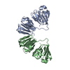 6fd8S S: Starting model for refinement C: citing same article ( |
|---|---|
| Similar structure data |


-Links
 PDBj
PDBj
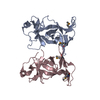
- Assembly
| Deposited unit | 
| ||||||||||||||||||||||||||||||||||||||||||||
|---|---|---|---|---|---|---|---|---|---|---|---|---|---|---|---|---|---|---|---|---|---|---|---|---|---|---|---|---|---|---|---|---|---|---|---|---|---|---|---|---|---|---|---|---|---|
| 1 |
| ||||||||||||||||||||||||||||||||||||||||||||
| Unit cell |
| ||||||||||||||||||||||||||||||||||||||||||||
| Noncrystallographic symmetry (NCS) | NCS domain:
NCS domain segments:
|
-Components
| #1: Protein | Mass: 20968.500 Da / Num. of mol.: 2 Source method: isolated from a genetically manipulated source Source: (gene. exp.) Homo sapiens (human) / Gene: CRYGS, CRYG8 / Production host:  Escherichia coli (E. coli) / References: UniProt: P22914 Escherichia coli (E. coli) / References: UniProt: P22914#2: Water | ChemComp-HOH / | Water Mass: 18.015 Da / Num. of mol.: 16 / Source method: isolated from a natural source / Formula: H2O Mass: 18.015 Da / Num. of mol.: 16 / Source method: isolated from a natural source / Formula: H2O |
|---|
-Experimental details
-Experiment
| Experiment | Method: X-RAY DIFFRACTION / Number of used crystals: 1 |
|---|
- Sample preparation
| Crystal | Density Matthews: 2.27 Å3/Da / Density % sol: 45.88 % |
|---|---|
| Crystal grow | Temperature: 293 K / Method: vapor diffusion Details: 50 mM HEPES pH 7.0, 20% PEG3350, 0.1 M sodium acetate pH 5.45 |
-Data collection
| Diffraction | Mean temperature: 293 K / Serial crystal experiment: Y |
|---|---|
| Diffraction source | Source: SYNCHROTRON / Site: PETRA III, EMBL c/o DESY  / Beamline: P14 (MX2) / Wavelength: 0.9762 Å / Beamline: P14 (MX2) / Wavelength: 0.9762 Å |
| Detector | Type: DECTRIS EIGER R 4M / Detector: PIXEL / Date: Jun 27, 2020 |
| Radiation | Protocol: SINGLE WAVELENGTH / Monochromatic (M) / Laue (L): M / Scattering type: x-ray |
| Radiation wavelength | Wavelength: 0.9762 Å / Relative weight: 1 |
| Reflection | Resolution: 3→69.6 Å / Num. obs: 8132 / % possible obs: 99.96 % / Redundancy: 48 % / Biso Wilson estimate: 52.32 Å2 / CC1/2: 0.9574 / Net I/σ(I): 2.5 |
| Reflection shell | Resolution: 3→3.108 Å / Num. unique obs: 787 / CC1/2: 0.885 |
| Serial crystallography sample delivery | Method: fixed target |
- Processing
| Software |
| ||||||||||||||||||||||||||||
|---|---|---|---|---|---|---|---|---|---|---|---|---|---|---|---|---|---|---|---|---|---|---|---|---|---|---|---|---|---|
| Refinement | Method to determine structure: MOLECULAR REPLACEMENT Starting model: 6FD8 Resolution: 3→69.6 Å / SU ML: 0.4305 / Cross valid method: FREE R-VALUE / σ(F): 1.34 / Phase error: 41.481 Stereochemistry target values: GeoStd + Monomer Library + CDL v1.2
| ||||||||||||||||||||||||||||
| Solvent computation | Shrinkage radii: 0.9 Å / VDW probe radii: 1.11 Å / Solvent model: FLAT BULK SOLVENT MODEL | ||||||||||||||||||||||||||||
| Displacement parameters | Biso mean: 52.64 Å2 | ||||||||||||||||||||||||||||
| Refinement step | Cycle: LAST / Resolution: 3→69.6 Å
| ||||||||||||||||||||||||||||
| Refine LS restraints |
| ||||||||||||||||||||||||||||
| Refine LS restraints NCS | Type: Torsion NCS / Rms dev position: 0.798069996017 Å | ||||||||||||||||||||||||||||
| LS refinement shell |
|
