structural constituent of eye lens / lens development in camera-type eye / visual perception / morphogenesis of an epithelium Similarity search - Function
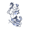
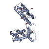
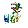
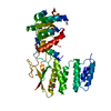
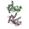
Journal: J Mol Biol / Year: 2019 Title: The Structure and Stability of the Disulfide-Linked γS-Crystallin Dimer Provide Insight into Oxidation Products Associated with Lens Cataract Formation. Authors: David C Thorn / Aidan B Grosas / Peter D Mabbitt / Nicholas J Ray / Colin J Jackson / John A Carver / Abstract: The reducing environment in the eye lens diminishes with age, leading to significant oxidative stress. Oxidation of lens crystallin proteins is the major contributor to their destabilization and ...The reducing environment in the eye lens diminishes with age, leading to significant oxidative stress. Oxidation of lens crystallin proteins is the major contributor to their destabilization and deleterious aggregation that scatters visible light, obscures vision, and ultimately leads to cataract. However, the molecular basis for oxidation-induced aggregation is unknown. Using X-ray crystallography and small-angle X-ray scattering, we describe the structure of a disulfide-linked dimer of human γS-crystallin that was obtained via oxidation of C24. The γS-crystallin dimer is stable at glutathione concentrations comparable to those in aged and cataractous lenses. Moreover, dimerization of γS-crystallin significantly increases the protein's propensity to form large insoluble aggregates owing to non-cooperative domain unfolding, as is observed in crystallin variants associated with early-onset cataract. These findings provide insight into how oxidative modification of crystallins contributes to cataract and imply that early-onset and age-related forms of the disease share comparable development pathways.
History
Deposition
Dec 22, 2017
Deposition site: PDBE / Processing site: PDBE
Revision 1.0
Dec 26, 2018
Provider: repository / Type: Initial release
Revision 1.1
Jan 16, 2019
Group: Data collection / Database references / Category: pdbx_database_related
In the structure databanks used in Yorodumi, some data are registered as the other names, "COVID-19 virus" and "2019-nCoV". Here are the details of the virus and the list of structure data.
Jan 31, 2019. EMDB accession codes are about to change! (news from PDBe EMDB page)
EMDB accession codes are about to change! (news from PDBe EMDB page)
The allocation of 4 digits for EMDB accession codes will soon come to an end. Whilst these codes will remain in use, new EMDB accession codes will include an additional digit and will expand incrementally as the available range of codes is exhausted. The current 4-digit format prefixed with “EMD-” (i.e. EMD-XXXX) will advance to a 5-digit format (i.e. EMD-XXXXX), and so on. It is currently estimated that the 4-digit codes will be depleted around Spring 2019, at which point the 5-digit format will come into force.
The EM Navigator/Yorodumi systems omit the EMD- prefix.
Related info.:Q: What is EMD? / ID/Accession-code notation in Yorodumi/EM Navigator
Yorodumi is a browser for structure data from EMDB, PDB, SASBDB, etc.
This page is also the successor to EM Navigator detail page, and also detail information page/front-end page for Omokage search.
The word "yorodu" (or yorozu) is an old Japanese word meaning "ten thousand". "mi" (miru) is to see.
Related info.:EMDB / PDB / SASBDB / Comparison of 3 databanks / Yorodumi Search / Aug 31, 2016. New EM Navigator & Yorodumi / Yorodumi Papers / Jmol/JSmol / Function and homology information / Changes in new EM Navigator and Yorodumi
 Movie
Movie Controller


 Open data
 Basic information
 Components
 Keywords
 STRUCTURAL PROTEIN /
  Function and homology information
 Authors
 Citation
 Structure visualization
 Downloads & links
 Other downloads
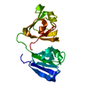


 PDBj
PDBj
 Assembly
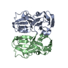


 Mass: 18.015 Da / Num. of mol.: 127 / Source method: isolated from a natural source / Formula: H2O
 Sample preparation
 Processing