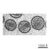
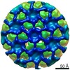
Journal: Commun Biol / Year: 2021 Title: CryoET structures of immature HIV Gag reveal six-helix bundle. Authors: Luiza Mendonça / Dapeng Sun / Jiying Ning / Jiwei Liu / Abhay Kotecha / Mateusz Olek / Thomas Frosio / Xiaofeng Fu / Benjamin A Himes / Alex B Kleinpeter / Eric O Freed / Jing Zhou / ...Authors: Luiza Mendonça / Dapeng Sun / Jiying Ning / Jiwei Liu / Abhay Kotecha / Mateusz Olek / Thomas Frosio / Xiaofeng Fu / Benjamin A Himes / Alex B Kleinpeter / Eric O Freed / Jing Zhou / Christopher Aiken / Peijun Zhang / Abstract: Gag is the HIV structural precursor protein which is cleaved by viral protease to produce mature infectious viruses. Gag is a polyprotein composed of MA (matrix), CA (capsid), SP1, NC (nucleocapsid), ...Gag is the HIV structural precursor protein which is cleaved by viral protease to produce mature infectious viruses. Gag is a polyprotein composed of MA (matrix), CA (capsid), SP1, NC (nucleocapsid), SP2 and p6 domains. SP1, together with the last eight residues of CA, have been hypothesized to form a six-helix bundle responsible for the higher-order multimerization of Gag necessary for HIV particle assembly. However, the structure of the complete six-helix bundle has been elusive. Here, we determined the structures of both Gag in vitro assemblies and Gag viral-like particles (VLPs) to 4.2 Å and 4.5 Å resolutions using cryo-electron tomography and subtomogram averaging by emClarity. A single amino acid mutation (T8I) in SP1 stabilizes the six-helix bundle, allowing to discern the entire CA-SP1 helix connecting to the NC domain. These structures provide a blueprint for future development of small molecule inhibitors that can lock SP1 in a stable helical conformation, interfere with virus maturation, and thus block HIV-1 infection.
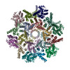
A: Gag protein B: Gag protein C: Gag protein D: Gag protein I: Gag protein N: Gag protein E: Gag protein J: Gag protein O: Gag protein F: Gag protein K: Gag protein P: Gag protein G: Gag protein L: Gag protein Q: Gag protein H: Gag protein M: Gag protein R: Gag protein
In the structure databanks used in Yorodumi, some data are registered as the other names, "COVID-19 virus" and "2019-nCoV". Here are the details of the virus and the list of structure data.
Jan 31, 2019. EMDB accession codes are about to change! (news from PDBe EMDB page)
EMDB accession codes are about to change! (news from PDBe EMDB page)
The allocation of 4 digits for EMDB accession codes will soon come to an end. Whilst these codes will remain in use, new EMDB accession codes will include an additional digit and will expand incrementally as the available range of codes is exhausted. The current 4-digit format prefixed with “EMD-” (i.e. EMD-XXXX) will advance to a 5-digit format (i.e. EMD-XXXXX), and so on. It is currently estimated that the 4-digit codes will be depleted around Spring 2019, at which point the 5-digit format will come into force.
The EM Navigator/Yorodumi systems omit the EMD- prefix.
Related info.:Q: What is EMD? / ID/Accession-code notation in Yorodumi/EM Navigator
Yorodumi is a browser for structure data from EMDB, PDB, SASBDB, etc.
This page is also the successor to EM Navigator detail page, and also detail information page/front-end page for Omokage search.
The word "yorodu" (or yorozu) is an old Japanese word meaning "ten thousand". "mi" (miru) is to see.
Related info.:EMDB / PDB / SASBDB / Comparison of 3 databanks / Yorodumi Search / Aug 31, 2016. New EM Navigator & Yorodumi / Yorodumi Papers / Jmol/JSmol / Function and homology information / Changes in new EM Navigator and Yorodumi
 Movie
Movie Controller


 Open data
 Basic information
 Components
 HIV-1 protease
  Keywords
 Function and homology information
 Authors
 United Kingdom, 1items
  Citation

 Structure visualization
 Downloads & links
 Other downloads


 PDBj
PDBj

 Assembly

 Sample preparation
 Electron microscopy imaging
 Processing