Movie
Movie Controller

[English] 日本語
 Yorodumi
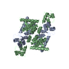
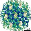
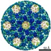
- PDB-6t61: A model of the EIAV CA-SP hexamer (C2) from Gag-deltaMA tubes ass... -
+ Open data

- Basic information
| Entry | Database: PDB / ID: 6t61 | |||||||||||||||||||||||||||||||||
|---|---|---|---|---|---|---|---|---|---|---|---|---|---|---|---|---|---|---|---|---|---|---|---|---|---|---|---|---|---|---|---|---|---|---|
| Title | A model of the EIAV CA-SP hexamer (C2) from Gag-deltaMA tubes assembled at pH8 | |||||||||||||||||||||||||||||||||
 Components Components | Gag polyprotein Group-specific antigen Group-specific antigen | |||||||||||||||||||||||||||||||||
 Keywords Keywords | VIRAL PROTEIN / Retrovirus / lentivirus / Equine infectious anemia virus / EIAV / Gag / capsid / IP6 / phytic acid / inositolhexakiphosphate | |||||||||||||||||||||||||||||||||
| Function / homology |  Function and homology information Function and homology informationviral budding via host ESCRT complex / viral nucleocapsid / structural constituent of virion / nucleic acid binding / zinc ion bindingSimilarity search - Function | |||||||||||||||||||||||||||||||||
| Biological species |  Equine infectious anemia virus Equine infectious anemia virus | |||||||||||||||||||||||||||||||||
| Method | ELECTRON MICROSCOPY / subtomogram averaging / cryo EM / Resolution: 3.7 Å | |||||||||||||||||||||||||||||||||
 Authors Authors | Dick, R.A. / Xu, C. / Morado, D.R. / Kravchuk, V. / Ricana, C.L. / Lyddon, T.D. / Broad, A.M. / Feathers, J.R. / Johnson, M.C. / Vogt, V.M. ...Dick, R.A. / Xu, C. / Morado, D.R. / Kravchuk, V. / Ricana, C.L. / Lyddon, T.D. / Broad, A.M. / Feathers, J.R. / Johnson, M.C. / Vogt, V.M. / Perilla, J.R. / Briggs, J.A.G. / Schur, F.K.M. | |||||||||||||||||||||||||||||||||
| Funding support |  Austria, Austria,  United States, United States,  United Kingdom, United Kingdom,  Germany, 10items Germany, 10items
| |||||||||||||||||||||||||||||||||
 Citation Citation | Journal: PLoS Pathog / Year: 2020 Title: Structures of immature EIAV Gag lattices reveal a conserved role for IP6 in lentivirus assembly. Authors: Robert A Dick / Chaoyi Xu / Dustin R Morado / Vladyslav Kravchuk / Clifton L Ricana / Terri D Lyddon / Arianna M Broad / J Ryan Feathers / Marc C Johnson / Volker M Vogt / Juan R Perilla / ...Authors: Robert A Dick / Chaoyi Xu / Dustin R Morado / Vladyslav Kravchuk / Clifton L Ricana / Terri D Lyddon / Arianna M Broad / J Ryan Feathers / Marc C Johnson / Volker M Vogt / Juan R Perilla / John A G Briggs / Florian K M Schur / Abstract: Retrovirus assembly is driven by the multidomain structural protein Gag. Interactions between the capsid domains (CA) of Gag result in Gag multimerization, leading to an immature virus particle that ...Retrovirus assembly is driven by the multidomain structural protein Gag. Interactions between the capsid domains (CA) of Gag result in Gag multimerization, leading to an immature virus particle that is formed by a protein lattice based on dimeric, trimeric, and hexameric protein contacts. Among retroviruses the inter- and intra-hexamer contacts differ, especially in the N-terminal sub-domain of CA (CANTD). For HIV-1 the cellular molecule inositol hexakisphosphate (IP6) interacts with and stabilizes the immature hexamer, and is required for production of infectious virus particles. We have used in vitro assembly, cryo-electron tomography and subtomogram averaging, atomistic molecular dynamics simulations and mutational analyses to study the HIV-related lentivirus equine infectious anemia virus (EIAV). In particular, we sought to understand the structural conservation of the immature lentivirus lattice and the role of IP6 in EIAV assembly. Similar to HIV-1, IP6 strongly promoted in vitro assembly of EIAV Gag proteins into virus-like particles (VLPs), which took three morphologically highly distinct forms: narrow tubes, wide tubes, and spheres. Structural characterization of these VLPs to sub-4Å resolution unexpectedly showed that all three morphologies are based on an immature lattice with preserved key structural components, highlighting the structural versatility of CA to form immature assemblies. A direct comparison between EIAV and HIV revealed that both lentiviruses maintain similar immature interfaces, which are established by both conserved and non-conserved residues. In both EIAV and HIV-1, IP6 regulates immature assembly via conserved lysine residues within the CACTD and SP. Lastly, we demonstrate that IP6 stimulates in vitro assembly of immature particles of several other retroviruses in the lentivirus genus, suggesting a conserved role for IP6 in lentiviral assembly. | |||||||||||||||||||||||||||||||||
| History |
|
- Structure visualization
| Movie |
Movie viewer |
|---|---|
| Structure viewer | Molecule: MolmilJmol/JSmol |
- Downloads & links
-Download
| PDBx/mmCIF format | 6t61.cif.gz | 703.1 KB | Display | PDBx/mmCIF format |
|---|---|---|---|---|
| PDB format | pdb6t61.ent.gz | 580.4 KB | Display | PDB format |
| PDBx/mmJSON format | 6t61.json.gz | Tree view | PDBx/mmJSON format | |
| Others |  Other downloads Other downloads |
-Validation report
| Arichive directory | https://data.pdbj.org/pub/pdb/validation_reports/t6/6t61ftp://data.pdbj.org/pub/pdb/validation_reports/t6/6t61 | HTTPS FTP |
|---|
-Related structure data
| Related structure data |  10381MC 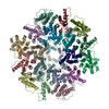 6t63C  6t64C C: citing same article ( M: map data used to model this data |
|---|---|
| Similar structure data |


-Links
 PDBj
PDBj
- Assembly
| Deposited unit | 
|
|---|---|
| 1 |
|
-Components
| #1: Protein | Group-specific antigen Mass: 54881.535 Da / Num. of mol.: 18 Source method: isolated from a genetically manipulated source Source: (gene. exp.) Equine infectious anemia virus / Gene: gag / Production host:  Escherichia coli BL21(DE3) (bacteria) / References: UniProt: P69730 Escherichia coli BL21(DE3) (bacteria) / References: UniProt: P69730 |
|---|
-Experimental details
-Experiment
| Experiment | Method: ELECTRON MICROSCOPY |
|---|---|
| EM experiment | Aggregation state: PARTICLE / 3D reconstruction method: subtomogram averaging |
- Sample preparation
| Component | Name: Equine infectious anemia virusEquine infectious anemia Type: VIRUS Details: Gag construct was expressed in E.coli and purified using the SUMO-tag system. Entity ID: all / Source: RECOMBINANT | ||||||||||||||||||||
|---|---|---|---|---|---|---|---|---|---|---|---|---|---|---|---|---|---|---|---|---|---|
| Molecular weight | Experimental value: NO | ||||||||||||||||||||
| Source (natural) | Organism: Equine infectious anemia virus | ||||||||||||||||||||
| Source (recombinant) | Organism: Escherichia coli BL21(DE3) (bacteria) | ||||||||||||||||||||
| Details of virus | Empty: YES / Enveloped: NO / Isolate: OTHER / Type: VIRUS-LIKE PARTICLE | ||||||||||||||||||||
| Natural host | Organism: Equus caballus | ||||||||||||||||||||
| Virus shell | Name: Capsid / Diameter: 700 nm | ||||||||||||||||||||
| Buffer solution | pH: 8 | ||||||||||||||||||||
| Buffer component |
| ||||||||||||||||||||
| Specimen | Embedding applied: NO / Shadowing applied: NO / Staining applied: NO / Vitrification applied: YES Details: Virus-like-particles (tubular) of EIAV Gag deltaMA-deltap9 (referred to as Gag deltaMA) assembled at pH8. | ||||||||||||||||||||
| Specimen support | Details: 20 mA / Grid material: COPPER / Grid mesh size: 300 divisions/in. / Grid type: C-flat-2/2 | ||||||||||||||||||||
| Vitrification | Instrument: FEI VITROBOT MARK II / Cryogen name: ETHANE / Humidity: 90 % / Chamber temperature: 288 K / Details: 1-2 seconds blot time, offset -3mm |
- Electron microscopy imaging
| Experimental equipment |  Model: Titan Krios / Image courtesy: FEI Company |
|---|---|
| Microscopy | Model: FEI TITAN KRIOS / Details: Nanoprobe |
| Electron gun | Electron source: FIELD EMISSION GUN / Accelerating voltage: 300 kV / Illumination mode: FLOOD BEAM |
| Electron lens | Mode: BRIGHT FIELDBright-field microscopy / Nominal magnification: 105000 X / Nominal defocus max: -5000 nm / Nominal defocus min: -1500 nm / Cs: 2.7 mm / C2 aperture diameter: 50 µm / Alignment procedure: ZEMLIN TABLEAU |
| Specimen holder | Cryogen: NITROGEN / Specimen holder model: FEI TITAN KRIOS AUTOGRID HOLDER |
| Image recording | Average exposure time: 1.8 sec. / Electron dose: 3.4 e/Å2 / Detector mode: SUPER-RESOLUTION / Film or detector model: GATAN K2 QUANTUM (4k x 4k) / Num. of grids imaged: 1 Details: Data was acquired using a dose-symmetric tilt acquisition scheme, as described in Hagen et al, 2017, J. Struct. Biol, 197(2):191-8 |
| EM imaging optics | Energyfilter name: GIF Quantum LS / Energyfilter slit width: 20 eV |
| Image scans | Width: 3708 / Height: 3838 / Movie frames/image: 20 |
- Processing
| EM software |
| ||||||||||||||||||||||||||||||||||||||||||||||||||||||||||||||||||||||
|---|---|---|---|---|---|---|---|---|---|---|---|---|---|---|---|---|---|---|---|---|---|---|---|---|---|---|---|---|---|---|---|---|---|---|---|---|---|---|---|---|---|---|---|---|---|---|---|---|---|---|---|---|---|---|---|---|---|---|---|---|---|---|---|---|---|---|---|---|---|---|---|
| Image processing | Details: Tilt series were low-pass filtered according to their cumulative dose using exposure filters that were calculated using an exposure-dependent amplitude attenuation function and critical ...Details: Tilt series were low-pass filtered according to their cumulative dose using exposure filters that were calculated using an exposure-dependent amplitude attenuation function and critical exposure constants (as published in Grant & Grigorieff, Elife, 2015). Tilt series were aligned and reconstructed in IMOD. | ||||||||||||||||||||||||||||||||||||||||||||||||||||||||||||||||||||||
| CTF correction | Details: CTF-correction was initially performed using ctfphaseflip in IMOD and NovaCTF in the final steps Type: PHASE FLIPPING AND AMPLITUDE CORRECTION | ||||||||||||||||||||||||||||||||||||||||||||||||||||||||||||||||||||||
| Symmetry | Point symmetry: C2 (2 fold cyclic) | ||||||||||||||||||||||||||||||||||||||||||||||||||||||||||||||||||||||
| 3D reconstruction | Resolution: 3.7 Å / Resolution method: FSC 0.143 CUT-OFF / Num. of particles: 89860 / Symmetry type: POINT | ||||||||||||||||||||||||||||||||||||||||||||||||||||||||||||||||||||||
| EM volume selection | Method: Subvolumes were defined according to their position on the VLPs Details: Subtomogram extraction positions were defined in Amira using the electron microscopy toolbox by determing the radii and the spline of the VLPs. Initially, positions were oversampled and ...Details: Subtomogram extraction positions were defined in Amira using the electron microscopy toolbox by determing the radii and the spline of the VLPs. Initially, positions were oversampled and subsequently cleaned during alignments using cross-correlation and distance thresholds. Num. of tomograms: 20 / Num. of volumes extracted: 308367 | ||||||||||||||||||||||||||||||||||||||||||||||||||||||||||||||||||||||
| Atomic model building | Protocol: OTHER / Space: REAL / Target criteria: Cross-correlation coefficient Details: Rigid body fitting was done in Chimera. Missing residues were built de novo in Coot. Refinement was performed iteratively in Phenix and Coot. | ||||||||||||||||||||||||||||||||||||||||||||||||||||||||||||||||||||||
| Atomic model building | PDB-ID: 2EIA |