 Movie
Movie Controller


+ Open data

- Basic information
| Entry | Database: PDB / ID: 6zph | |||||||||||||||||||||
|---|---|---|---|---|---|---|---|---|---|---|---|---|---|---|---|---|---|---|---|---|---|---|
| Title | Kinesin binding protein complexed with Kif15 motor domain | |||||||||||||||||||||
 Components Components |
| |||||||||||||||||||||
 Keywords Keywords |  MOTOR PROTEIN / Kinesin / microtubules / kinesin binding protein / KBP MOTOR PROTEIN / Kinesin / microtubules / kinesin binding protein / KBP | |||||||||||||||||||||
| Function / homology |  Function and homology information Function and homology informationplus-end kinesin complex / central nervous system projection neuron axonogenesis / Kinesins / COPI-dependent Golgi-to-ER retrograde traffic / kinesin complex / microtubule motor activity / mitochondrial transport / microtubule-based movement / cytoskeletal motor activity / kinesin binding ...plus-end kinesin complex / central nervous system projection neuron axonogenesis / Kinesins / COPI-dependent Golgi-to-ER retrograde traffic / kinesin complex / microtubule motor activity / mitochondrial transport / microtubule-based movement / cytoskeletal motor activity / kinesin binding / MHC class II antigen presentation / neuron projection maintenance / spindle / microtubule cytoskeleton organization / mitotic cell cycle / microtubule binding / in utero embryonic development / microtubule / cytoskeleton / centrosome / ATP hydrolysis activity / mitochondrion / ATP binding / membrane / cytosolSimilarity search - Function | |||||||||||||||||||||
| Biological species |  Homo sapiens (human) Homo sapiens (human) | |||||||||||||||||||||
| Method | ELECTRON MICROSCOPY / single particle reconstruction / cryo EM / Resolution: 6.9 Å | |||||||||||||||||||||
 Authors Authors | Atherton, J. / Hummel, J.J.A. / Olieric, N. / Locke, J. / Pena, A. / Rosenfeld, S.S. / Steinmetz, M.O. / Hoogenraad, C.C. / Moores, C.A. | |||||||||||||||||||||
| Funding support |  United Kingdom, United Kingdom,  Switzerland, Switzerland,  United States, 6items United States, 6items
| |||||||||||||||||||||
 Citation Citation | Journal: Elife / Year: 2020 Title: The mechanism of kinesin inhibition by kinesin-binding protein. Authors: Joseph Atherton / Jessica Ja Hummel / Natacha Olieric / Julia Locke / Alejandro Peña / Steven S Rosenfeld / Michel O Steinmetz / Casper C Hoogenraad / Carolyn A Moores /  Abstract: Subcellular compartmentalisation is necessary for eukaryotic cell function. Spatial and temporal regulation of kinesin activity is essential for building these local environments via control of ...Subcellular compartmentalisation is necessary for eukaryotic cell function. Spatial and temporal regulation of kinesin activity is essential for building these local environments via control of intracellular cargo distribution. Kinesin-binding protein (KBP) interacts with a subset of kinesins via their motor domains, inhibits their microtubule (MT) attachment, and blocks their cellular function. However, its mechanisms of inhibition and selectivity have been unclear. Here we use cryo-electron microscopy to reveal the structure of KBP and of a KBP-kinesin motor domain complex. KBP is a tetratricopeptide repeat-containing, right-handed α-solenoid that sequesters the kinesin motor domain's tubulin-binding surface, structurally distorting the motor domain and sterically blocking its MT attachment. KBP uses its α-solenoid concave face and edge loops to bind the kinesin motor domain, and selected structure-guided mutations disrupt KBP inhibition of kinesin transport in cells. The KBP-interacting motor domain surface contains motifs exclusively conserved in KBP-interacting kinesins, suggesting a basis for kinesin selectivity. | |||||||||||||||||||||
| History |
|
- Structure visualization
| Movie |
Movie viewer |
|---|---|
| Structure viewer | Molecule: MolmilJmol/JSmol |
- Downloads & links
-Download
| PDBx/mmCIF format | 6zph.cif.gz | 154.8 KB | Display | PDBx/mmCIF format |
|---|---|---|---|---|
| PDB format | pdb6zph.ent.gz | 117.6 KB | Display | PDB format |
| PDBx/mmJSON format | 6zph.json.gz | Tree view | PDBx/mmJSON format | |
| Others |  Other downloads Other downloads |
-Validation report
| Arichive directory | https://data.pdbj.org/pub/pdb/validation_reports/zp/6zphftp://data.pdbj.org/pub/pdb/validation_reports/zp/6zph | HTTPS FTP |
|---|
-Related structure data
| Related structure data | 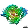 11339MC 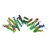 6zpgC  6zpiC C: citing same article ( M: map data used to model this data |
|---|---|
| Similar structure data |


-Links
 PDBj
PDBj


- Assembly
| Deposited unit | 
|
|---|---|
| 1 |
|
-Components
| #1: Protein | Mass: 71913.945 Da / Num. of mol.: 1 Source method: isolated from a genetically manipulated source Source: (gene. exp.) Homo sapiens (human) / Gene: KIFBP, KBP, KIAA1279, KIF1BP / Production host:  Escherichia coli (E. coli) / References: UniProt: Q96EK5 Escherichia coli (E. coli) / References: UniProt: Q96EK5 |
|---|---|
| #2: Protein | Mass: 41986.004 Da / Num. of mol.: 1 Source method: isolated from a genetically manipulated source Source: (gene. exp.) Homo sapiens (human) / Gene: KIF15, KLP2, KNSL7 / Production host: Escherichia coli (E. coli) / References: UniProt: Q9NS87 |
| #3: Chemical | ChemComp-ADP / Adenosine diphosphate  Mass: 427.201 Da / Num. of mol.: 1 / Source method: obtained synthetically / Formula: C10H15N5O10P2 / Comment: ADP, energy-carrying molecule*YM Mass: 427.201 Da / Num. of mol.: 1 / Source method: obtained synthetically / Formula: C10H15N5O10P2 / Comment: ADP, energy-carrying molecule*YM |
| #4: Chemical | ChemComp-MG /   Mass: 24.305 Da / Num. of mol.: 1 / Source method: obtained synthetically / Formula: Mg Mass: 24.305 Da / Num. of mol.: 1 / Source method: obtained synthetically / Formula: Mg |
| Has ligand of interest | N |
-Experimental details
-Experiment
| Experiment | Method: ELECTRON MICROSCOPY |
|---|---|
| EM experiment | Aggregation state: PARTICLE / 3D reconstruction method: single particle reconstruction |
- Sample preparation
| Component | Name: Kinesin binding protein complexed with Kif15 motor domain cryo-EM density Type: COMPLEX / Entity ID: #1-#2 / Source: RECOMBINANT |
|---|---|
| Molecular weight | Value: 0.072 MDa / Experimental value: YES |
| Source (natural) | Organism: Homo sapiens (human) |
| Source (recombinant) | Organism: Escherichia coli (E. coli) |
| Buffer solution | pH: 7.5 |
| Specimen | Embedding applied: NO / Shadowing applied: NO / Staining applied: NO / Vitrification applied: YES |
| Vitrification | Cryogen name: ETHANE |
- Electron microscopy imaging
| Experimental equipment |  Model: Titan Krios / Image courtesy: FEI Company |
|---|---|
| Microscopy | Model: FEI TITAN KRIOS |
| Electron gun | Electron source: FIELD EMISSION GUN / Accelerating voltage: 300 kV / Illumination mode: FLOOD BEAM |
| Electron lens | Mode: BRIGHT FIELDBright-field microscopy |
| Image recording | Electron dose: 42 e/Å2 / Detector mode: COUNTING / Film or detector model: GATAN K2 SUMMIT (4k x 4k) / Details: Movies were dose weighted. |
- Processing
| Software | Name: PHENIX / Version: 1.14_3260: / Classification: refinement |
|---|---|
| EM software | Name: RELION / Category: final Euler assignment |
| CTF correction | Type: PHASE FLIPPING AND AMPLITUDE CORRECTION |
| 3D reconstruction | Resolution: 6.9 Å / Resolution method: FSC 0.143 CUT-OFF / Num. of particles: 7513 / Symmetry type: POINT |
