 Movie
Movie Controller

[English] 日本語
 Yorodumi
- PDB-6y5y: Structure of New Jersey Polyomavirus VP1 in complex with 3'-Sialy... -
+ Open data

- Basic information
| Entry | Database: PDB / ID: 6y5y | |||||||||
|---|---|---|---|---|---|---|---|---|---|---|
| Title | Structure of New Jersey Polyomavirus VP1 in complex with 3'-Sialyllactose | |||||||||
 Components Components | VP1 | |||||||||
 Keywords Keywords |  VIRAL PROTEIN / Major Capsid Protein / Polyomavirus VIRAL PROTEIN / Major Capsid Protein / Polyomavirus | |||||||||
| Function / homology | Capsid protein VP1,Polyomavirus / Polyomavirus capsid protein VP1 superfamily / Polyomavirus coat protein / Double-stranded DNA virus, group I, capsid / viral capsid / structural molecule activity / 3'-sialyl-alpha-lactose / VP1 Function and homology information Function and homology information | |||||||||
| Biological species |  New Jersey polyomavirus-2013 New Jersey polyomavirus-2013 | |||||||||
| Method | X-RAY DIFFRACTION / SYNCHROTRON / MOLECULAR REPLACEMENT / Resolution: 1.8 Å | |||||||||
 Authors Authors | Stroh, L.J. / Rustmeier, N.H. / Stehle, T. | |||||||||
| Funding support |  Germany, 1items Germany, 1items
| |||||||||
 Citation Citation | Journal: Mbio / Year: 2020 Title: Structural Basis and Evolution of Glycan Receptor Specificities within the Polyomavirus Family. Authors: Stroh, L.J. / Rustmeier, N.H. / Blaum, B.S. / Botsch, J. / Rossler, P. / Wedekink, F. / Lipkin, W.I. / Mishra, N. / Stehle, T. | |||||||||
| History |
|
- Structure visualization
| Structure viewer | Molecule: MolmilJmol/JSmol |
|---|
- Downloads & links
-Download
| PDBx/mmCIF format | 6y5y.cif.gz | 767.5 KB | Display | PDBx/mmCIF format |
|---|---|---|---|---|
| PDB format | pdb6y5y.ent.gz | 507.8 KB | Display | PDB format |
| PDBx/mmJSON format | 6y5y.json.gz | Tree view | PDBx/mmJSON format | |
| Others |  Other downloads Other downloads |
-Validation report
| Arichive directory | https://data.pdbj.org/pub/pdb/validation_reports/y5/6y5yftp://data.pdbj.org/pub/pdb/validation_reports/y5/6y5y | HTTPS FTP |
|---|
-Related structure data
| Related structure data | 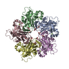 6y5xC 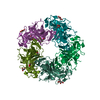 6y5zC 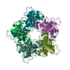 6y60C 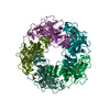 6y61C  6y63C  6y64C  6y65C  6y66C  6y67C  6y6aC  6y9iC  4fmgS S: Starting model for refinement C: citing same article ( |
|---|---|
| Similar structure data |


-Links
 PDBj
PDBj


- Assembly
| Deposited unit | 
| ||||||||||||
|---|---|---|---|---|---|---|---|---|---|---|---|---|---|
| 1 | 
| ||||||||||||
| 2 | 
| ||||||||||||
| Unit cell |
|
-Components
-Protein , 1 types, 10 molecules ABCDEFGHIJ
| #1: Protein | Mass: 34096.566 Da / Num. of mol.: 10 Source method: isolated from a genetically manipulated source Source: (gene. exp.) New Jersey polyomavirus-2013 / Gene: VP1 / Production host:  Escherichia coli BL21(DE3) (bacteria) / Strain (production host): Rosetta 2 / References: UniProt: A0A024B5J2 Escherichia coli BL21(DE3) (bacteria) / Strain (production host): Rosetta 2 / References: UniProt: A0A024B5J2 |
|---|
-Sugars , 2 types, 10 molecules
| #2: Polysaccharide | N-acetyl-alpha-neuraminic acid-(2-3)-beta-D-galactopyranose-(1-4)-alpha-D-glucopyranose / 3'-sialyl-alpha-lactose   , Oligosaccharide / Class: Nutrient / Mass: 633.552 Da / Num. of mol.: 4 , Oligosaccharide / Class: Nutrient / Mass: 633.552 Da / Num. of mol.: 4Source method: isolated from a genetically manipulated source Details: oligosaccharide / References: 3'-sialyl-alpha-lactose #3: Polysaccharide | N-acetyl-alpha-neuraminic acid-(2-3)-beta-D-galactopyranose-(1-4)-beta-D-glucopyranose / Mass: 633.552 Da / Num. of mol.: 6Source method: isolated from a genetically manipulated source |
|---|
-Non-polymers , 3 types, 2826 molecules 


| #4: Chemical | ChemComp-GOL / Glycerol Mass: 92.094 Da / Num. of mol.: 39 / Source method: obtained synthetically / Formula: C3H8O3 Mass: 92.094 Da / Num. of mol.: 39 / Source method: obtained synthetically / Formula: C3H8O3#5: Chemical | ChemComp-MG /  Mass: 24.305 Da / Num. of mol.: 10 / Source method: obtained synthetically / Formula: Mg Mass: 24.305 Da / Num. of mol.: 10 / Source method: obtained synthetically / Formula: Mg#6: Water | ChemComp-HOH / | WaterMass: 18.015 Da / Num. of mol.: 2777 / Source method: isolated from a natural source / Formula: H2O |
|---|
-Details
| Has ligand of interest | Y |
|---|
-Experimental details
-Experiment
| Experiment | Method: X-RAY DIFFRACTION / Number of used crystals: 1 |
|---|
- Sample preparation
| Crystal | Density Matthews: 2.4 Å3/Da / Density % sol: 48.66 % |
|---|---|
| Crystal grow | Temperature: 277 K / Method: vapor diffusion, sitting drop / pH: 7 / Details: Succinic acid, PEG 3350 |
-Data collection
| Diffraction | Mean temperature: 93 K / Serial crystal experiment: N |
|---|---|
| Diffraction source | Source: SYNCHROTRON / Site: BESSY / Beamline: 14.1 / Wavelength: 0.918409 Å |
| Detector | Type: DECTRIS PILATUS 6M / Detector: PIXEL / Date: Sep 19, 2014 |
| Radiation | Monochromator: DCM Si(111) / Protocol: SINGLE WAVELENGTH / Monochromatic (M) / Laue (L): M / Scattering type: x-ray |
| Radiation wavelength | Wavelength: 0.918409 Å / Relative weight: 1 |
| Reflection | Resolution: 1.8→48.2 Å / Num. obs: 295112 / % possible obs: 99.83 % / Redundancy: 5.7 % / Biso Wilson estimate: 19.03 Å2 / CC1/2: 0.996 / Rrim(I) all: 0.1556 / Net I/σ(I): 9.79 |
| Reflection shell | Resolution: 1.801→1.865 Å / Mean I/σ(I) obs: 1.39 / Num. unique obs: 29218 / CC1/2: 0.603 / Rrim(I) all: 1.176 |
- Processing
| Software |
| |||||||||||||||||||||||||||||||||||||||||||||||||||||||||||||||||||||||||||||||||||||||||||||||||||||||||||||||||||||||||||||||||||||||||||||||||||||||||||||||||||||||||||||||||||||||||||||||||||||||||||||||||||||||||
|---|---|---|---|---|---|---|---|---|---|---|---|---|---|---|---|---|---|---|---|---|---|---|---|---|---|---|---|---|---|---|---|---|---|---|---|---|---|---|---|---|---|---|---|---|---|---|---|---|---|---|---|---|---|---|---|---|---|---|---|---|---|---|---|---|---|---|---|---|---|---|---|---|---|---|---|---|---|---|---|---|---|---|---|---|---|---|---|---|---|---|---|---|---|---|---|---|---|---|---|---|---|---|---|---|---|---|---|---|---|---|---|---|---|---|---|---|---|---|---|---|---|---|---|---|---|---|---|---|---|---|---|---|---|---|---|---|---|---|---|---|---|---|---|---|---|---|---|---|---|---|---|---|---|---|---|---|---|---|---|---|---|---|---|---|---|---|---|---|---|---|---|---|---|---|---|---|---|---|---|---|---|---|---|---|---|---|---|---|---|---|---|---|---|---|---|---|---|---|---|---|---|---|---|---|---|---|---|---|---|---|---|---|---|---|---|---|---|---|
| Refinement | Method to determine structure: MOLECULAR REPLACEMENT Starting model: 4FMG Resolution: 1.8→48.2 Å / SU ML: 0.2213 / Cross valid method: FREE R-VALUE / σ(F): 1.35 / Phase error: 21.455
| |||||||||||||||||||||||||||||||||||||||||||||||||||||||||||||||||||||||||||||||||||||||||||||||||||||||||||||||||||||||||||||||||||||||||||||||||||||||||||||||||||||||||||||||||||||||||||||||||||||||||||||||||||||||||
| Solvent computation | Shrinkage radii: 0.9 Å / VDW probe radii: 1.11 Å | |||||||||||||||||||||||||||||||||||||||||||||||||||||||||||||||||||||||||||||||||||||||||||||||||||||||||||||||||||||||||||||||||||||||||||||||||||||||||||||||||||||||||||||||||||||||||||||||||||||||||||||||||||||||||
| Displacement parameters | Biso mean: 23.67 Å2 | |||||||||||||||||||||||||||||||||||||||||||||||||||||||||||||||||||||||||||||||||||||||||||||||||||||||||||||||||||||||||||||||||||||||||||||||||||||||||||||||||||||||||||||||||||||||||||||||||||||||||||||||||||||||||
| Refinement step | Cycle: LAST / Resolution: 1.8→48.2 Å
| |||||||||||||||||||||||||||||||||||||||||||||||||||||||||||||||||||||||||||||||||||||||||||||||||||||||||||||||||||||||||||||||||||||||||||||||||||||||||||||||||||||||||||||||||||||||||||||||||||||||||||||||||||||||||
| Refine LS restraints |
| |||||||||||||||||||||||||||||||||||||||||||||||||||||||||||||||||||||||||||||||||||||||||||||||||||||||||||||||||||||||||||||||||||||||||||||||||||||||||||||||||||||||||||||||||||||||||||||||||||||||||||||||||||||||||
| LS refinement shell |
|
