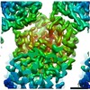
 Movie
Movie Controller


+ Open data

- Basic information
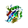
| Entry | Database: PDB / ID: 6u5g | |||||||||||||||
|---|---|---|---|---|---|---|---|---|---|---|---|---|---|---|---|---|
| Title | MicroED structure of a FIB-milled CypA Crystal | |||||||||||||||
 Components Components | Peptidyl-prolyl cis-trans isomerase A | |||||||||||||||
 Keywords Keywords |  ISOMERASE / Peptidyl-prolyl / cis-trans / cyclophilin ISOMERASE / Peptidyl-prolyl / cis-trans / cyclophilin | |||||||||||||||
| Function / homology |  Function and homology information Function and homology informationnegative regulation of protein K48-linked ubiquitination / negative regulation of viral life cycle / regulation of apoptotic signaling pathway / cell adhesion molecule production / lipid droplet organization / heparan sulfate binding / regulation of viral genome replication / leukocyte chemotaxis / negative regulation of stress-activated MAPK cascade / endothelial cell activation ...negative regulation of protein K48-linked ubiquitination / negative regulation of viral life cycle / regulation of apoptotic signaling pathway / cell adhesion molecule production / lipid droplet organization / heparan sulfate binding / regulation of viral genome replication / leukocyte chemotaxis / negative regulation of stress-activated MAPK cascade / endothelial cell activation / virion binding / Basigin interactions / cyclosporin A binding / Minus-strand DNA synthesis / Plus-strand DNA synthesis / Uncoating of the HIV Virion / Early Phase of HIV Life Cycle / Integration of provirus / APOBEC3G mediated resistance to HIV-1 infection / Calcineurin activates NFAT / viral release from host cell / Binding and entry of HIV virion / positive regulation of viral genome replication / protein peptidyl-prolyl isomerization / negative regulation of oxidative stress-induced intrinsic apoptotic signaling pathway / positive regulation of protein dephosphorylation / Gene and protein expression by JAK-STAT signaling after Interleukin-12 stimulation / activation of protein kinase B activity / neutrophil chemotaxis / negative regulation of protein phosphorylation / peptidylprolyl isomerase / peptidyl-prolyl cis-trans isomerase activity / positive regulation of protein secretion / Assembly Of The HIV Virion / negative regulation of protein kinase activity / Budding and maturation of HIV virion / neuron differentiation / platelet activation / platelet aggregation / SARS-CoV-1 activates/modulates innate immune responses / unfolded protein binding / integrin binding / protein folding / Platelet degranulation / cellular response to oxidative stress / positive regulation of NF-kappaB transcription factor activity / secretory granule lumen / vesicle / ficolin-1-rich granule lumen / positive regulation of MAPK cascade / response to hypoxia / positive regulation of protein phosphorylation / focal adhesion / apoptotic process / Neutrophil degranulation / protein-containing complex / extracellular space / RNA binding / extracellular exosome / extracellular region / membrane / nucleus / cytosol / cytoplasmSimilarity search - Function | |||||||||||||||
| Biological species |  Homo sapiens (human) Homo sapiens (human) | |||||||||||||||
| Method | ELECTRON CRYSTALLOGRAPHY / electron crystallography / MOLECULAR REPLACEMENT / cryo EM / Resolution: 2.5 Å | |||||||||||||||
 Authors Authors | Wolff, A.M. / Martynowycz, M.W. / Zhao, W. / Gonen, T. / Fraser, J.S. / Thompson, M.C. | |||||||||||||||
| Funding support |  United States, 4items United States, 4items
| |||||||||||||||
 Citation Citation | Journal: IUCrJ / Year: 2020 Title: Comparing serial X-ray crystallography and microcrystal electron diffraction (MicroED) as methods for routine structure determination from small macromolecular crystals. Authors: Alexander M Wolff / Iris D Young / Raymond G Sierra / Aaron S Brewster / Michael W Martynowycz / Eriko Nango / Michihiro Sugahara / Takanori Nakane / Kazutaka Ito / Andrew Aquila / Asmit ...Authors: Alexander M Wolff / Iris D Young / Raymond G Sierra / Aaron S Brewster / Michael W Martynowycz / Eriko Nango / Michihiro Sugahara / Takanori Nakane / Kazutaka Ito / Andrew Aquila / Asmit Bhowmick / Justin T Biel / Sergio Carbajo / Aina E Cohen / Saul Cortez / Ana Gonzalez / Tomoya Hino / Dohyun Im / Jake D Koralek / Minoru Kubo / Tomas S Lazarou / Takashi Nomura / Shigeki Owada / Avi J Samelson / Tomoyuki Tanaka / Rie Tanaka / Erin M Thompson / Henry van den Bedem / Rahel A Woldeyes / Fumiaki Yumoto / Wei Zhao / Kensuke Tono / Sebastien Boutet / So Iwata / Tamir Gonen / Nicholas K Sauter / James S Fraser / Michael C Thompson /  Abstract: Innovative new crystallographic methods are facilitating structural studies from ever smaller crystals of biological macromolecules. In particular, serial X-ray crystallography and microcrystal ...Innovative new crystallographic methods are facilitating structural studies from ever smaller crystals of biological macromolecules. In particular, serial X-ray crystallography and microcrystal electron diffraction (MicroED) have emerged as useful methods for obtaining structural information from crystals on the nanometre to micrometre scale. Despite the utility of these methods, their implementation can often be difficult, as they present many challenges that are not encountered in traditional macromolecular crystallography experiments. Here, XFEL serial crystallography experiments and MicroED experiments using batch-grown microcrystals of the enzyme cyclophilin A are described. The results provide a roadmap for researchers hoping to design macromolecular microcrystallography experiments, and they highlight the strengths and weaknesses of the two methods. Specifically, we focus on how the different physical conditions imposed by the sample-preparation and delivery methods required for each type of experiment affect the crystal structure of the enzyme. | |||||||||||||||
| History |
|
- Structure visualization
| Movie |
Movie viewer |
|---|---|
| Structure viewer | Molecule: MolmilJmol/JSmol |
- Downloads & links
-Download
| PDBx/mmCIF format | 6u5g.cif.gz | 87.5 KB | Display | PDBx/mmCIF format |
|---|---|---|---|---|
| PDB format | pdb6u5g.ent.gz | 52 KB | Display | PDB format |
| PDBx/mmJSON format | 6u5g.json.gz | Tree view | PDBx/mmJSON format | |
| Others |  Other downloads Other downloads |
-Validation report
| Arichive directory | https://data.pdbj.org/pub/pdb/validation_reports/u5/6u5gftp://data.pdbj.org/pub/pdb/validation_reports/u5/6u5g | HTTPS FTP |
|---|
-Related structure data


-Links
 PDBj
PDBj


- Assembly
| Deposited unit | 
| ||||||||||||
|---|---|---|---|---|---|---|---|---|---|---|---|---|---|
| 1 |
| ||||||||||||
| Unit cell |
|
-Components
| #1: Protein | Mass: 18036.504 Da / Num. of mol.: 1 Source method: isolated from a genetically manipulated source Source: (gene. exp.) Homo sapiens (human) / Gene: PPIA, CYPA / Production host:  Escherichia coli BL21(DE3) (bacteria) / References: UniProt: P62937, peptidylprolyl isomerase Escherichia coli BL21(DE3) (bacteria) / References: UniProt: P62937, peptidylprolyl isomerase |
|---|---|
| #2: Water | ChemComp-HOH / Water Mass: 18.015 Da / Num. of mol.: 32 / Source method: isolated from a natural source / Formula: H2O Mass: 18.015 Da / Num. of mol.: 32 / Source method: isolated from a natural source / Formula: H2O |
-Experimental details
-Experiment
| Experiment | Method: ELECTRON CRYSTALLOGRAPHY |
|---|---|
| EM experiment | Aggregation state: 3D ARRAY / 3D reconstruction method: electron crystallography |
- Sample preparation
| Component | Name: Peptidyl-prolyl cis-trans isomerase A / Type: COMPLEX / Details: monomer / Entity ID: #1 / Source: RECOMBINANT |
|---|---|
| Source (natural) | Organism: Homo sapiens (human) |
| Source (recombinant) | Organism: Escherichia coli BL21(DE3) (bacteria) |
| EM crystal formation | Details: 600 uL of protein at 60 mg/mL was combined with 400 uL of 50 percent PEG 3350 in a glass vial and stirred with an Octagon stir bar at 500 RPM Lipid mixture: NA / Temperature: 296 K |
| Buffer solution | pH: 7.5 |
| Specimen | Embedding applied: NO / Shadowing applied: NO / Staining applied: NO / Vitrification applied: YES |
| Specimen support | Details: unspecified |
| Vitrification | Cryogen name: ETHANE |
| Crystal | Density Matthews: 2.75 Å3/Da / Density % sol: 55.34 % |
-Data collection
| Experimental equipment | 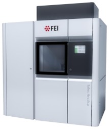 Model: Talos Arctica / Image courtesy: FEI Company | ||||||||||||||||||||||||||||
|---|---|---|---|---|---|---|---|---|---|---|---|---|---|---|---|---|---|---|---|---|---|---|---|---|---|---|---|---|---|
| Microscopy | Model: FEI TALOS ARCTICA | ||||||||||||||||||||||||||||
| Electron gun | Electron source: FIELD EMISSION GUN / Accelerating voltage: 200 kV / Illumination mode: OTHER | ||||||||||||||||||||||||||||
| Electron lens | Mode: DIFFRACTION | ||||||||||||||||||||||||||||
| Image recording | Electron dose: 0.06 e/Å2 / Film or detector model: TVIPS TEMCAM-F416 (4k x 4k) / Num. of grids imaged: 1 | ||||||||||||||||||||||||||||
| EM diffraction | Camera length: 2055 mm | ||||||||||||||||||||||||||||
| EM diffraction shell |
| ||||||||||||||||||||||||||||
| EM diffraction stats | Fourier space coverage: 86 % / High resolution: 2.5 Å / Num. of intensities measured: 22370 / Num. of structure factors: 6236 / Phase error: 23.06 ° / Phase residual: 23.06 ° / Phase error rejection criteria: 0 / Rmerge: 0.217 / Rsym: 0.217 | ||||||||||||||||||||||||||||
| Reflection | Biso Wilson estimate: 35.52 Å2 |
- Processing
| Software |
| ||||||||||||||||||||||||||||
|---|---|---|---|---|---|---|---|---|---|---|---|---|---|---|---|---|---|---|---|---|---|---|---|---|---|---|---|---|---|
| EM software |
| ||||||||||||||||||||||||||||
| EM 3D crystal entity | ∠α: 90 ° / ∠β: 90 ° / ∠γ: 90 ° / A: 42.4 Å / B: 53.4 Å / C: 87.76 Å / Space group name: P212121 / Space group num: 19 | ||||||||||||||||||||||||||||
| CTF correction | Type: NONE | ||||||||||||||||||||||||||||
| 3D reconstruction | Resolution: 2.5 Å / Resolution method: DIFFRACTION PATTERN/LAYERLINES / Symmetry type: 3D CRYSTAL | ||||||||||||||||||||||||||||
| Atomic model building | Space: RECIPROCAL | ||||||||||||||||||||||||||||
| Atomic model building | PDB-ID: 4YUM Accession code: 4YUM / Source name: PDB / Type: experimental model | ||||||||||||||||||||||||||||
| Refinement | Method to determine structure: MOLECULAR REPLACEMENT Starting model: PDB entry 4YUM Resolution: 2.5→26.48 Å / SU ML: 0.4224 / Cross valid method: FREE R-VALUE / σ(F): 1.34 / Phase error: 22.5393
| ||||||||||||||||||||||||||||
| Solvent computation | Shrinkage radii: 0.9 Å / VDW probe radii: 1.11 Å | ||||||||||||||||||||||||||||
| Displacement parameters | Biso mean: 30.6 Å2 | ||||||||||||||||||||||||||||
| Refine LS restraints |
| ||||||||||||||||||||||||||||
| LS refinement shell |
|
