 Movie
Movie Controller


+ Open data

- Basic information
| Entry | Database: PDB / ID: 6thm | ||||||
|---|---|---|---|---|---|---|---|
| Title | Linalool Dehydratase Isomerase M125A mutant | ||||||
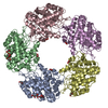
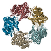
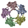
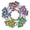
 Components Components | Linalool dehydratase-isomerase protein LDI | ||||||
 Keywords Keywords |  LYASE / Alkene / Alkenol / Hydratase LYASE / Alkene / Alkenol / Hydratase | ||||||
| Function / homology |  Function and homology informationlinalool dehydratase / geraniol isomerase / monoterpene catabolic process / intramolecular hydroxytransferase activity / monoterpenoid metabolic process / hydro-lyase activity / cellular response to organic substance / protein tetramerization / periplasmic space Function and homology informationlinalool dehydratase / geraniol isomerase / monoterpene catabolic process / intramolecular hydroxytransferase activity / monoterpenoid metabolic process / hydro-lyase activity / cellular response to organic substance / protein tetramerization / periplasmic spaceSimilarity search - Function | ||||||
| Biological species |  Castellaniella defragrans 65Phen (bacteria) Castellaniella defragrans 65Phen (bacteria) | ||||||
| Method | X-RAY DIFFRACTION / SYNCHROTRON / MOLECULAR REPLACEMENT / Resolution: 1.99 Å | ||||||
 Authors Authors | Cuetos, A. / Zukic, E. / Danesh-Azari, H.R. / Grogan, G. | ||||||
| Funding support |  United Kingdom, 1items United Kingdom, 1items
| ||||||
 Citation Citation | Journal: Acs Catalysis / Year: 2020 Title: Mutational Analysis of Linalool Dehydratase Isomerase Suggests That Alcohol and Alkene Transformations Are Catalyzed Using Noncovalent Mechanisms Authors: Cuetos, A. / Iglesias-Fernandez, J. / Danesh-Azari, H.R. / Zukic, E. / Dowle, A. / Osuna, S. / Grogan, G. | ||||||
| History |
|
- Structure visualization
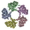
| Structure viewer | Molecule: MolmilJmol/JSmol |
|---|
- Downloads & links
-Download
| PDBx/mmCIF format | 6thm.cif.gz | 374.9 KB | Display | PDBx/mmCIF format |
|---|---|---|---|---|
| PDB format | pdb6thm.ent.gz | 306.3 KB | Display | PDB format |
| PDBx/mmJSON format | 6thm.json.gz | Tree view | PDBx/mmJSON format | |
| Others |  Other downloads Other downloads |
-Validation report
| Arichive directory | https://data.pdbj.org/pub/pdb/validation_reports/th/6thmftp://data.pdbj.org/pub/pdb/validation_reports/th/6thm | HTTPS FTP |
|---|
-Related structure data
| Related structure data | 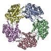 6t9hC 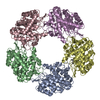 6tfnC  6tfrC 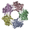 6tftC 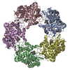 5g1uS S: Starting model for refinement C: citing same article ( |
|---|---|
| Similar structure data |


-Links
 PDBj
PDBj- Assembly
| Deposited unit | 
| |||||||||
|---|---|---|---|---|---|---|---|---|---|---|
| 1 |
| |||||||||
| Unit cell |
| |||||||||
| Components on special symmetry positions |
|
-Components
| #1: Protein | Mass: 41933.598 Da / Num. of mol.: 5 Source method: isolated from a genetically manipulated source Details: M125A mutant Source: (gene. exp.) Castellaniella defragrans 65Phen (bacteria)Gene: BN940_14136 / Production host: Escherichia coli BL21(DE3) (bacteria) / References: UniProt: W8X534#2: Chemical | ChemComp-EDO / Ethylene glycol  Mass: 62.068 Da / Num. of mol.: 15 / Source method: obtained synthetically / Formula: C2H6O2 Mass: 62.068 Da / Num. of mol.: 15 / Source method: obtained synthetically / Formula: C2H6O2#3: Chemical | Malonic acid  Mass: 102.046 Da / Num. of mol.: 2 / Source method: obtained synthetically / Formula: C3H2O4 Mass: 102.046 Da / Num. of mol.: 2 / Source method: obtained synthetically / Formula: C3H2O4#4: Water | ChemComp-HOH / | Water Mass: 18.015 Da / Num. of mol.: 824 / Source method: isolated from a natural source / Formula: H2O Mass: 18.015 Da / Num. of mol.: 824 / Source method: isolated from a natural source / Formula: H2OHas ligand of interest | N | |
|---|
-Experimental details
-Experiment
| Experiment | Method: X-RAY DIFFRACTION / Number of used crystals: 1 |
|---|
- Sample preparation
| Crystal | Density Matthews: 2.82 Å3/Da / Density % sol: 56.39 % |
|---|---|
| Crystal grow | Temperature: 298 K / Method: vapor diffusion, hanging drop / pH: 7.5 Details: 0.2 M sodium malonate; 0.1 M bis-tris propane pH 7.5; 20% (w/w) PEG3350; 5% (v/v) methylpentanediol |
-Data collection
| Diffraction | Mean temperature: 120 K / Serial crystal experiment: N |
|---|---|
| Diffraction source | Source: SYNCHROTRON / Site: Diamond / Beamline: I03 / Wavelength: 0.97625 Å |
| Detector | Type: DECTRIS EIGER X 16M / Detector: PIXEL / Date: May 20, 2018 |
| Radiation | Protocol: SINGLE WAVELENGTH / Monochromatic (M) / Laue (L): M / Scattering type: x-ray |
| Radiation wavelength | Wavelength: 0.97625 Å / Relative weight: 1 |
| Reflection | Resolution: 1.99→98.33 Å / Num. obs: 156439 / % possible obs: 98.4 % / Redundancy: 4.2 % / CC1/2: 1 / Rmerge(I) obs: 0.08 / Rpim(I) all: 0.07 / Net I/σ(I): 9.1 |
| Reflection shell | Resolution: 1.99→2.02 Å / Rmerge(I) obs: 0.91 / Mean I/σ(I) obs: 1.6 / Num. unique obs: 7677 / CC1/2: 0.68 / Rpim(I) all: 0.77 |
- Processing
| Software |
| ||||||||||||||||||||||||||||||||||||||||||||||||||||||||||||
|---|---|---|---|---|---|---|---|---|---|---|---|---|---|---|---|---|---|---|---|---|---|---|---|---|---|---|---|---|---|---|---|---|---|---|---|---|---|---|---|---|---|---|---|---|---|---|---|---|---|---|---|---|---|---|---|---|---|---|---|---|---|
| Refinement | Method to determine structure: MOLECULAR REPLACEMENT Starting model: 5G1U Resolution: 1.99→96.18 Å / Cor.coef. Fo:Fc: 0.965 / Cor.coef. Fo:Fc free: 0.947 / SU B: 4.662 / SU ML: 0.122 / Cross valid method: THROUGHOUT / σ(F): 0 / ESU R: 0.152 / ESU R Free: 0.141 Details: HYDROGENS HAVE BEEN ADDED IN THE RIDING POSITIONS U VALUES : REFINED INDIVIDUALLY
| ||||||||||||||||||||||||||||||||||||||||||||||||||||||||||||
| Solvent computation | Ion probe radii: 0.8 Å / Shrinkage radii: 0.8 Å / VDW probe radii: 1.2 Å | ||||||||||||||||||||||||||||||||||||||||||||||||||||||||||||
| Displacement parameters | Biso max: 95.42 Å2 / Biso mean: 33.191 Å2 / Biso min: 21.02 Å2
| ||||||||||||||||||||||||||||||||||||||||||||||||||||||||||||
| Refinement step | Cycle: final / Resolution: 1.99→96.18 Å
| ||||||||||||||||||||||||||||||||||||||||||||||||||||||||||||
| Refine LS restraints |
| ||||||||||||||||||||||||||||||||||||||||||||||||||||||||||||
| LS refinement shell | Resolution: 1.99→2.042 Å / Rfactor Rfree error: 0 / Total num. of bins used: 20
|
