Movie
Movie Controller

[English] 日本語
 Yorodumi
- PDB-6t1r: Pseudo-atomic model of a 16-mer assembly of reduced recombinant h... -
+ Open data

- Basic information
| Entry | Database: PDB / ID: 6t1r | |||||||||
|---|---|---|---|---|---|---|---|---|---|---|
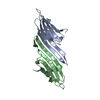
| Title | Pseudo-atomic model of a 16-mer assembly of reduced recombinant human alphaA-crystallin (non domain swapped configuration) | |||||||||
 Components Components | Alpha-crystallin A chain | |||||||||
 Keywords Keywords |  CHAPERONE / sHsp / alphaA-crystallin / domain swapping CHAPERONE / sHsp / alphaA-crystallin / domain swapping | |||||||||
| Function / homology |  Function and homology information Function and homology informationnegative regulation of intracellular transport / structural constituent of eye lens / response to stimulus / visual perception / unfolded protein binding / protein stabilization / negative regulation of apoptotic process / structural molecule activity / protein-containing complex / nucleoplasm ...negative regulation of intracellular transport / structural constituent of eye lens / response to stimulus / visual perception / unfolded protein binding / protein stabilization / negative regulation of apoptotic process / structural molecule activity / protein-containing complex / nucleoplasm / identical protein binding / metal ion binding / nucleus / cytosol / cytoplasmSimilarity search - Function | |||||||||
| Biological species |  Homo sapiens (human) Homo sapiens (human) | |||||||||
| Method | ELECTRON MICROSCOPY / single particle reconstruction / cryo EM / Resolution: 9.8 Å | |||||||||
 Authors Authors | Peters, C. / Kaiser, C.J.O. / Weinkauf, S. / Zacharias, M. / Buchner, J. | |||||||||
| Funding support |  Germany, 2items Germany, 2items
| |||||||||
 Citation Citation | Journal: Nat Struct Mol Biol / Year: 2019 Title: The structure and oxidation of the eye lens chaperone αA-crystallin. Authors: Christoph J O Kaiser / Carsten Peters / Philipp W N Schmid / Maria Stavropoulou / Juan Zou / Vinay Dahiya / Evgeny V Mymrikov / Beate Rockel / Sam Asami / Martin Haslbeck / Juri Rappsilber / ...Authors: Christoph J O Kaiser / Carsten Peters / Philipp W N Schmid / Maria Stavropoulou / Juan Zou / Vinay Dahiya / Evgeny V Mymrikov / Beate Rockel / Sam Asami / Martin Haslbeck / Juri Rappsilber / Bernd Reif / Martin Zacharias / Johannes Buchner / Sevil Weinkauf /  Abstract: The small heat shock protein αA-crystallin is a molecular chaperone important for the optical properties of the vertebrate eye lens. It forms heterogeneous oligomeric ensembles. We determined the ...The small heat shock protein αA-crystallin is a molecular chaperone important for the optical properties of the vertebrate eye lens. It forms heterogeneous oligomeric ensembles. We determined the structures of human αA-crystallin oligomers by combining cryo-electron microscopy, cross-linking/mass spectrometry, NMR spectroscopy and molecular modeling. The different oligomers can be interconverted by the addition or subtraction of tetramers, leading to mainly 12-, 16- and 20-meric assemblies in which interactions between N-terminal regions are important. Cross-dimer domain-swapping of the C-terminal region is a determinant of αA-crystallin heterogeneity. Human αA-crystallin contains two cysteines, which can form an intramolecular disulfide in vivo. Oxidation in vitro requires conformational changes and oligomer dissociation. The oxidized oligomers, which are larger than reduced αA-crystallin and destabilized against unfolding, are active chaperones and can transfer the disulfide to destabilized substrate proteins. The insight into the structure and function of αA-crystallin provides a basis for understanding its role in the eye lens. | |||||||||
| History |
|
- Structure visualization
| Movie |
Movie viewer |
|---|---|
| Structure viewer | Molecule: MolmilJmol/JSmol |
- Downloads & links
-Download
| PDBx/mmCIF format | 6t1r.cif.gz | 375.8 KB | Display | PDBx/mmCIF format |
|---|---|---|---|---|
| PDB format | pdb6t1r.ent.gz | 258.5 KB | Display | PDB format |
| PDBx/mmJSON format | 6t1r.json.gz | Tree view | PDBx/mmJSON format | |
| Others |  Other downloads Other downloads |
-Validation report
| Arichive directory | https://data.pdbj.org/pub/pdb/validation_reports/t1/6t1rftp://data.pdbj.org/pub/pdb/validation_reports/t1/6t1r | HTTPS FTP |
|---|
-Related structure data
| Related structure data | 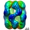 4894MC 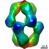 4895C  4896C M: map data used to model this data C: citing same article ( |
|---|---|
| Similar structure data |
-Links
 PDBj
PDBj


- Assembly
| Deposited unit | 
|
|---|---|
| 1 |
|
-Components
| #1: Protein | Mass: 19936.314 Da / Num. of mol.: 16 Source method: isolated from a genetically manipulated source Details: wild type residues 1-166 / Source: (gene. exp.) Homo sapiens (human) / Tissue: lens / Gene: CRYAA, CRYA1, HSPB4 / Organ: eye / Plasmid: pET28 / Production host:  Escherichia coli BL21 (bacteria) / Variant (production host): CodonPlus-RIL / References: UniProt: P02489 Escherichia coli BL21 (bacteria) / Variant (production host): CodonPlus-RIL / References: UniProt: P02489 |
|---|
-Experimental details
-Experiment
| Experiment | Method: ELECTRON MICROSCOPY |
|---|---|
| EM experiment | Aggregation state: PARTICLE / 3D reconstruction method: single particle reconstruction |
- Sample preparation
| Component | Name: Reduced recombinant human alphaA-crystallin / Type: COMPLEX Details: Recombinant wild type full-length human alphaA-crystallin purified in the presence of reductant. Entity ID: all / Source: RECOMBINANT | |||||||||||||||||||||||||||||||||||
|---|---|---|---|---|---|---|---|---|---|---|---|---|---|---|---|---|---|---|---|---|---|---|---|---|---|---|---|---|---|---|---|---|---|---|---|---|
| Molecular weight | Value: 0.319 MDa / Experimental value: NO | |||||||||||||||||||||||||||||||||||
| Source (natural) | Organism: Homo sapiens (human) / Cellular location: cytoplasm / Organ: eye / Tissue: lens | |||||||||||||||||||||||||||||||||||
| Source (recombinant) | Organism: Escherichia coli BL21 (bacteria) / Plasmid: pET28 | |||||||||||||||||||||||||||||||||||
| Buffer solution | pH: 7.4 Details: Buffer was prepared without EDTA and DTT. EDTA stock (500mM) was titrated to pH 8 and added to 1 mM. DTT stock (1M) was added to 1mM. | |||||||||||||||||||||||||||||||||||
| Buffer component |
| |||||||||||||||||||||||||||||||||||
| Specimen | Conc.: 0.3 mg/ml / Embedding applied: NO / Shadowing applied: NO / Staining applied: NO / Vitrification applied: YES Details: Specimen was thawed, diluted to the final concentration and equilibrated at 310K for 3h. | |||||||||||||||||||||||||||||||||||
| Specimen support | Grid material: COPPER / Grid mesh size: 200 divisions/in. / Grid type: Quantifoil R2/1 | |||||||||||||||||||||||||||||||||||
| Vitrification | Instrument: HOMEMADE PLUNGER / Cryogen name: ETHANE / Chamber temperature: 293 K Details: Diluted equilibrated specimen was added to glow-discharged (30s) grids. Sample was blotted 30s after sample application and immediately plunged. |
- Electron microscopy imaging
| Experimental equipment |  Model: Titan Krios / Image courtesy: FEI Company |
|---|---|
| Microscopy | Model: FEI TITAN KRIOS |
| Electron gun | Electron source: FIELD EMISSION GUN / Accelerating voltage: 300 kV / Illumination mode: FLOOD BEAM |
| Electron lens | Mode: BRIGHT FIELDBright-field microscopy / Nominal magnification: 37000 X / Calibrated magnification: 37037 X / Nominal defocus max: 2500 nm / Nominal defocus min: 1200 nm / Cs: 2.7 mm / C2 aperture diameter: 70 µm / Alignment procedure: COMA FREE |
| Specimen holder | Cryogen: NITROGEN / Specimen holder model: FEI TITAN KRIOS AUTOGRID HOLDER |
| Image recording | Average exposure time: 3.2 sec. / Electron dose: 30 e/Å2 / Detector mode: SUPER-RESOLUTION / Film or detector model: GATAN K2 SUMMIT (4k x 4k) / Num. of grids imaged: 3 |
| EM imaging optics | Energyfilter slit width: 10 eV |
| Image scans | Movie frames/image: 10 / Used frames/image: 1-10 |
- Processing
| EM software |
| ||||||||||||||||||||||||||||
|---|---|---|---|---|---|---|---|---|---|---|---|---|---|---|---|---|---|---|---|---|---|---|---|---|---|---|---|---|---|
| CTF correction | Type: PHASE FLIPPING ONLY | ||||||||||||||||||||||||||||
| Particle selection | Num. of particles selected: 74068 / Details: particles were picked manually | ||||||||||||||||||||||||||||
| Symmetry | Point symmetry: D4 (2x4 fold dihedral) | ||||||||||||||||||||||||||||
| 3D reconstruction | Resolution: 9.8 Å / Resolution method: FSC 0.143 CUT-OFF / Num. of particles: 19783 / Symmetry type: POINT | ||||||||||||||||||||||||||||
| Atomic model building | Protocol: FLEXIBLE FIT Details: Structure created by homology modelling using PDB 3N3E and the N-terminal region (1-60) modelled using I-Tasser. The model was refined by flexible fitting into the EM density map EMD-4894. ...Details: Structure created by homology modelling using PDB 3N3E and the N-terminal region (1-60) modelled using I-Tasser. The model was refined by flexible fitting into the EM density map EMD-4894. The N-terminus is modelled only as CA atoms. | ||||||||||||||||||||||||||||
| Atomic model building | PDB-ID: 3N3E Accession code: 3N3E / Source name: PDB / Type: experimental model |