 Movie
Movie Controller


+ Open data

- Basic information
| Entry | Database: PDB / ID: 3n3e | ||||||
|---|---|---|---|---|---|---|---|
| Title | Zebrafish AlphaA crystallin | ||||||
 Components Components | Alpha A crystallin | ||||||
 Keywords Keywords |  CHAPERONE / protein chaperone / eye lens transparency CHAPERONE / protein chaperone / eye lens transparency | ||||||
| Function / homology |  Function and homology informationeye development / structural constituent of eye lens / lens development in camera-type eye / visual perception / unfolded protein binding / metal ion binding / cytoplasm Function and homology informationeye development / structural constituent of eye lens / lens development in camera-type eye / visual perception / unfolded protein binding / metal ion binding / cytoplasmSimilarity search - Function | ||||||
| Biological species |  Danio rerio (zebrafish) Danio rerio (zebrafish) | ||||||
| Method | X-RAY DIFFRACTION / SYNCHROTRON / MOLECULAR REPLACEMENT / molecular replacement / Resolution: 1.75 Å | ||||||
 Authors Authors | Laganowsky, A. / Eisenberg, D. | ||||||
 Citation Citation | Journal: Protein Sci. / Year: 2010 Title: Non-3D domain swapped crystal structure of truncated zebrafish alphaA crystallin. Authors: Laganowsky, A. / Eisenberg, D. | ||||||
| History |
|
- Structure visualization
| Structure viewer | Molecule: MolmilJmol/JSmol |
|---|
- Downloads & links
-Download
| PDBx/mmCIF format | 3n3e.cif.gz | 100.9 KB | Display | PDBx/mmCIF format |
|---|---|---|---|---|
| PDB format | pdb3n3e.ent.gz | 78.6 KB | Display | PDB format |
| PDBx/mmJSON format | 3n3e.json.gz | Tree view | PDBx/mmJSON format | |
| Others |  Other downloads Other downloads |
-Validation report
| Arichive directory | https://data.pdbj.org/pub/pdb/validation_reports/n3/3n3eftp://data.pdbj.org/pub/pdb/validation_reports/n3/3n3e | HTTPS FTP |
|---|
-Related structure data
| Related structure data | 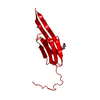 3l1eS S: Starting model for refinement |
|---|---|
| Similar structure data |


-Links
 PDBj
PDBj


- Assembly
| Deposited unit | 
| ||||||||
|---|---|---|---|---|---|---|---|---|---|
| 1 |
| ||||||||
| Unit cell |
|
-Components
| #1: Protein | Mass: 11959.220 Da / Num. of mol.: 2 / Fragment: UNP residues 61- 166 Source method: isolated from a genetically manipulated source Source: (gene. exp.) Danio rerio (zebrafish) / Gene: cryaa / Plasmid: pET28 / Production host:  Escherichia coli (E. coli) / Strain (production host): BL21(DE3) / References: UniProt: Q8UUZ6 Escherichia coli (E. coli) / Strain (production host): BL21(DE3) / References: UniProt: Q8UUZ6#2: Water | ChemComp-HOH / | Water Mass: 18.015 Da / Num. of mol.: 171 / Source method: isolated from a natural source / Formula: H2O Mass: 18.015 Da / Num. of mol.: 171 / Source method: isolated from a natural source / Formula: H2O |
|---|
-Experimental details
-Experiment
| Experiment | Method: X-RAY DIFFRACTION / Number of used crystals: 2 |
|---|
- Sample preparation
| Crystal | Density Matthews: 2.05 Å3/Da / Density % sol: 40.09 % |
|---|---|
| Crystal grow | Temperature: 298 K / Method: vapor diffusion, hanging drop / pH: 6 Details: 0.1M MIB pH 6.0, 25% PEG 1500, VAPOR DIFFUSION, HANGING DROP, temperature 298K |
-Data collection
| Diffraction | Mean temperature: 100 K | ||||||||||||||||||||||||||||||||||||||||||||||||||||||||||||||||||
|---|---|---|---|---|---|---|---|---|---|---|---|---|---|---|---|---|---|---|---|---|---|---|---|---|---|---|---|---|---|---|---|---|---|---|---|---|---|---|---|---|---|---|---|---|---|---|---|---|---|---|---|---|---|---|---|---|---|---|---|---|---|---|---|---|---|---|---|
| Diffraction source | Source: SYNCHROTRON / Site: APS  / Beamline: 24-ID-E / Wavelength: 0.979 Å / Beamline: 24-ID-E / Wavelength: 0.979 Å | ||||||||||||||||||||||||||||||||||||||||||||||||||||||||||||||||||
| Detector | Type: ADSC QUANTUM 315 / Detector: CCD / Date: Feb 20, 2010 | ||||||||||||||||||||||||||||||||||||||||||||||||||||||||||||||||||
| Radiation | Protocol: SINGLE WAVELENGTH / Monochromatic (M) / Laue (L): M / Scattering type: x-ray | ||||||||||||||||||||||||||||||||||||||||||||||||||||||||||||||||||
| Radiation wavelength | Wavelength: 0.979 Å / Relative weight: 1 | ||||||||||||||||||||||||||||||||||||||||||||||||||||||||||||||||||
| Reflection | Resolution: 1.75→30 Å / Num. obs: 21407 / % possible obs: 98.2 % / Redundancy: 7 % / Rmerge(I) obs: 0.063 / Χ2: 1.046 / Net I/σ(I): 11.4 | ||||||||||||||||||||||||||||||||||||||||||||||||||||||||||||||||||
| Reflection shell |
|
-Phasing
| Phasing | Method: molecular replacement |
|---|
- Processing
| Software |
| |||||||||||||||||||||||||||||||||||||||||||||||||||||||||||||||
|---|---|---|---|---|---|---|---|---|---|---|---|---|---|---|---|---|---|---|---|---|---|---|---|---|---|---|---|---|---|---|---|---|---|---|---|---|---|---|---|---|---|---|---|---|---|---|---|---|---|---|---|---|---|---|---|---|---|---|---|---|---|---|---|---|
| Refinement | Method to determine structure: MOLECULAR REPLACEMENT Starting model: PDB entry 3L1E Resolution: 1.75→26.615 Å / Occupancy max: 1 / Occupancy min: 0.25 / FOM work R set: 0.839 / SU ML: 0.22 / σ(F): 0.06 / Stereochemistry target values: ML
| |||||||||||||||||||||||||||||||||||||||||||||||||||||||||||||||
| Solvent computation | Shrinkage radii: 0.9 Å / VDW probe radii: 1.11 Å / Solvent model: FLAT BULK SOLVENT MODEL / Bsol: 29.133 Å2 / ksol: 0.342 e/Å3 | |||||||||||||||||||||||||||||||||||||||||||||||||||||||||||||||
| Displacement parameters | Biso max: 74.52 Å2 / Biso mean: 24.739 Å2 / Biso min: 10.1 Å2
| |||||||||||||||||||||||||||||||||||||||||||||||||||||||||||||||
| Refinement step | Cycle: LAST / Resolution: 1.75→26.615 Å
| |||||||||||||||||||||||||||||||||||||||||||||||||||||||||||||||
| Refine LS restraints |
| |||||||||||||||||||||||||||||||||||||||||||||||||||||||||||||||
| LS refinement shell | Refine-ID: X-RAY DIFFRACTION / Total num. of bins used: 8
|
