 Movie
Movie Controller


+ Open data

- Basic information
| Entry | Database: PDB / ID: 6s5d | |||||||||
|---|---|---|---|---|---|---|---|---|---|---|
| Title | Square conformation of KtrA R16A mutant ring with bound ATP | |||||||||
 Components Components | Ktr system potassium uptake protein A | |||||||||
 Keywords Keywords |  TRANSPORT PROTEIN / RCK domain / potassium homeostasis / cation channel / magnesium / square conformation octameric ring / atp TRANSPORT PROTEIN / RCK domain / potassium homeostasis / cation channel / magnesium / square conformation octameric ring / atp | |||||||||
| Function / homology |  Function and homology information Function and homology informationmonoatomic cation transmembrane transporter activity / potassium ion transport / identical protein binding / plasma membraneSimilarity search - Function | |||||||||
| Biological species |  Bacillus subtilis subsp. subtilis str. 168 (bacteria) Bacillus subtilis subsp. subtilis str. 168 (bacteria) | |||||||||
| Method | X-RAY DIFFRACTION / SYNCHROTRON / MOLECULAR REPLACEMENT / Resolution: 3.393 Å | |||||||||
 Authors Authors | Teixeira-Duarte, C.M. / Fonseca, F. / Morais-Cabral, J.H. | |||||||||
| Funding support |  Portugal, 2items Portugal, 2items
| |||||||||
 Citation Citation | Journal: Elife / Year: 2019 Title: Activation of a nucleotide-dependent RCK domain requires binding of a cation cofactor to a conserved site. Authors: Teixeira-Duarte, C.M. / Fonseca, F. / Morais Cabral, J.H. | |||||||||
| History |
|
- Structure visualization
| Structure viewer | Molecule: MolmilJmol/JSmol |
|---|
- Downloads & links
-Download
| PDBx/mmCIF format | 6s5d.cif.gz | 102.7 KB | Display | PDBx/mmCIF format |
|---|---|---|---|---|
| PDB format | pdb6s5d.ent.gz | 76.5 KB | Display | PDB format |
| PDBx/mmJSON format | 6s5d.json.gz | Tree view | PDBx/mmJSON format | |
| Others |  Other downloads Other downloads |
-Validation report
| Arichive directory | https://data.pdbj.org/pub/pdb/validation_reports/s5/6s5dftp://data.pdbj.org/pub/pdb/validation_reports/s5/6s5d | HTTPS FTP |
|---|
-Related structure data
| Related structure data | 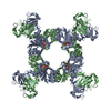 6s2jC 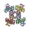 6s5bC 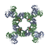 6s5cC 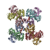 6s5eC 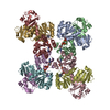 6s5gC 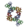 6s5nC 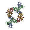 6s5oC 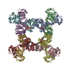 6s7rC 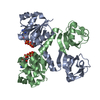 4j90S C: citing same article ( S: Starting model for refinement |
|---|---|
| Similar structure data |


-Links
 PDBj
PDBj
- Assembly
| Deposited unit | 
| ||||||||
|---|---|---|---|---|---|---|---|---|---|
| 1 | 
| ||||||||
| Unit cell |
|
-Components
| #1: Protein | Mass: 24830.645 Da / Num. of mol.: 2 / Mutation: R16A Source method: isolated from a genetically manipulated source Source: (gene. exp.) Bacillus subtilis subsp. subtilis str. 168 (bacteria)Gene: ktrA, yuaA, BSU31090 / Production host: Escherichia coli (E. coli) / References: UniProt: O32080#2: Chemical | Adenosine triphosphate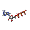  Mass: 507.181 Da / Num. of mol.: 2 / Source method: obtained synthetically / Formula: C10H16N5O13P3 / Feature type: SUBJECT OF INVESTIGATION / Comment: ATP, energy-carrying molecule*YM Mass: 507.181 Da / Num. of mol.: 2 / Source method: obtained synthetically / Formula: C10H16N5O13P3 / Feature type: SUBJECT OF INVESTIGATION / Comment: ATP, energy-carrying molecule*YM#3: Chemical | ChemComp-MG / |   Mass: 24.305 Da / Num. of mol.: 1 / Source method: obtained synthetically / Formula: Mg / Feature type: SUBJECT OF INVESTIGATION Mass: 24.305 Da / Num. of mol.: 1 / Source method: obtained synthetically / Formula: Mg / Feature type: SUBJECT OF INVESTIGATION#4: Water | ChemComp-HOH / | Water Mass: 18.015 Da / Num. of mol.: 2 / Source method: isolated from a natural source / Formula: H2O Mass: 18.015 Da / Num. of mol.: 2 / Source method: isolated from a natural source / Formula: H2OHas ligand of interest | Y | |
|---|
-Experimental details
-Experiment
| Experiment | Method: X-RAY DIFFRACTION / Number of used crystals: 1 |
|---|
- Sample preparation
| Crystal | Density Matthews: 3.11 Å3/Da / Density % sol: 60.51 % |
|---|---|
| Crystal grow | Temperature: 293.15 K / Method: vapor diffusion / pH: 7.5 Details: 100mM HEPES-NaOH pH 7.5, 2% PEG 6000, 1% 2-Methyl-2,4-pentanediol (MPD) |
-Data collection
| Diffraction | Mean temperature: 100 K / Serial crystal experiment: N |
|---|---|
| Diffraction source | Source: SYNCHROTRON / Site: SOLEIL  / Beamline: PROXIMA 1 / Wavelength: 0.97857 Å / Beamline: PROXIMA 1 / Wavelength: 0.97857 Å |
| Detector | Type: DECTRIS PILATUS 6M / Detector: PIXEL / Date: Dec 6, 2014 |
| Radiation | Protocol: SINGLE WAVELENGTH / Monochromatic (M) / Laue (L): M / Scattering type: x-ray |
| Radiation wavelength | Wavelength: 0.97857 Å / Relative weight: 1 |
| Reflection | Resolution: 3.39→45.6 Å / Num. obs: 8535 / % possible obs: 99.8 % / Redundancy: 8.9 % / CC1/2: 1 / Rmerge(I) obs: 0.047 / Rpim(I) all: 0.017 / Rrim(I) all: 0.05 / Net I/σ(I): 17.1 |
| Reflection shell | Resolution: 3.39→3.66 Å / Redundancy: 8.5 % / Rmerge(I) obs: 1.829 / Mean I/σ(I) obs: 1.3 / Num. unique obs: 1724 / CC1/2: 0.573 / Rpim(I) all: 0.655 / % possible all: 99 |
- Processing
| Software |
| |||||||||||||||||||||||||||||||||||||||||||||||||||||||||||||||||||||||||||||||||||||||||||
|---|---|---|---|---|---|---|---|---|---|---|---|---|---|---|---|---|---|---|---|---|---|---|---|---|---|---|---|---|---|---|---|---|---|---|---|---|---|---|---|---|---|---|---|---|---|---|---|---|---|---|---|---|---|---|---|---|---|---|---|---|---|---|---|---|---|---|---|---|---|---|---|---|---|---|---|---|---|---|---|---|---|---|---|---|---|---|---|---|---|---|---|---|
| Refinement | Method to determine structure: MOLECULAR REPLACEMENT Starting model: 4j90 Resolution: 3.393→45.598 Å / SU ML: 0.82 / Cross valid method: FREE R-VALUE / σ(F): 1.3 / Phase error: 41.08
| |||||||||||||||||||||||||||||||||||||||||||||||||||||||||||||||||||||||||||||||||||||||||||
| Solvent computation | Shrinkage radii: 0.9 Å / VDW probe radii: 1.11 Å | |||||||||||||||||||||||||||||||||||||||||||||||||||||||||||||||||||||||||||||||||||||||||||
| Refinement step | Cycle: LAST / Resolution: 3.393→45.598 Å
| |||||||||||||||||||||||||||||||||||||||||||||||||||||||||||||||||||||||||||||||||||||||||||
| Refine LS restraints |
| |||||||||||||||||||||||||||||||||||||||||||||||||||||||||||||||||||||||||||||||||||||||||||
| LS refinement shell |
|
