 Movie
Movie Controller


+ Open data

- Basic information
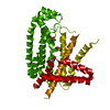
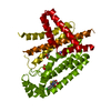
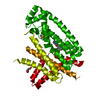
| Entry | Database: PDB / ID: 6r36 | ||||||
|---|---|---|---|---|---|---|---|
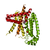
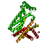
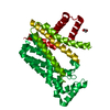
| Title | T. brucei farnesyl pyrophosphate synthase (FPPS) | ||||||
 Components Components | Farnesyl pyrophosphate synthase Dimethylallyltranstransferase Dimethylallyltranstransferase | ||||||
 Keywords Keywords | TRANSFERASE / sterol biosynthesis / farnesyl diphosphate synthase / Trypanosoma brucei / homodimer | ||||||
| Function / homology |  Function and homology information Function and homology informationtransferase activity, transferring alkyl or aryl (other than methyl) groups / isoprenoid biosynthetic process / metal ion bindingSimilarity search - Function | ||||||
| Biological species |  Trypanosoma brucei (eukaryote) Trypanosoma brucei (eukaryote) | ||||||
| Method | X-RAY DIFFRACTION / SYNCHROTRON / MOLECULAR REPLACEMENT / molecular replacement / Resolution: 1.67 Å | ||||||
 Authors Authors | Muenzker, L. / Petrick, J.K. / Schleberger, C. / Jahnke, W. | ||||||
| Funding support |  Switzerland, 1items Switzerland, 1items
| ||||||
 Citation Citation | Journal: Chembiochem / Year: 2020 Title: Fragment-Based Discovery of Non-bisphosphonate Binders of Trypanosoma brucei Farnesyl Pyrophosphate Synthase. Authors: Munzker, L. / Petrick, J.K. / Schleberger, C. / Clavel, D. / Cornaciu, I. / Wilcken, R. / Marquez, J.A. / Klebe, G. / Marzinzik, A. / Jahnke, W. | ||||||
| History |
|
- Structure visualization
| Structure viewer | Molecule: MolmilJmol/JSmol |
|---|
- Downloads & links
-Download
| PDBx/mmCIF format | 6r36.cif.gz | 147.8 KB | Display | PDBx/mmCIF format |
|---|---|---|---|---|
| PDB format | pdb6r36.ent.gz | 115.8 KB | Display | PDB format |
| PDBx/mmJSON format | 6r36.json.gz | Tree view | PDBx/mmJSON format | |
| Others |  Other downloads Other downloads |
-Validation report
| Arichive directory | https://data.pdbj.org/pub/pdb/validation_reports/r3/6r36ftp://data.pdbj.org/pub/pdb/validation_reports/r3/6r36 | HTTPS FTP |
|---|
-Related structure data
| Related structure data | 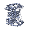 4rypS S: Starting model for refinement |
|---|---|
| Similar structure data |


-Links
 PDBj
PDBj
- Assembly
| Deposited unit | 
| ||||||||
|---|---|---|---|---|---|---|---|---|---|
| 1 | 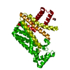
| ||||||||
| Unit cell |
| ||||||||
| Components on special symmetry positions |
|
-Components
| #1: Protein | Dimethylallyltranstransferase Mass: 42169.211 Da / Num. of mol.: 1 Source method: isolated from a genetically manipulated source Details: GP is an expression tag / Source: (gene. exp.) Trypanosoma brucei (eukaryote) / Production host:  Escherichia coli BL21(DE3) (bacteria) / References: UniProt: Q86C09 Escherichia coli BL21(DE3) (bacteria) / References: UniProt: Q86C09 | ||||
|---|---|---|---|---|---|
| #2: Chemical | Dimethyl sulfoxide  Mass: 78.133 Da / Num. of mol.: 2 / Source method: obtained synthetically / Formula: C2H6OS / Comment: DMSO, precipitant*YM Mass: 78.133 Da / Num. of mol.: 2 / Source method: obtained synthetically / Formula: C2H6OS / Comment: DMSO, precipitant*YM#3: Chemical | ChemComp-PEG / | Diethylene glycol  Mass: 106.120 Da / Num. of mol.: 1 / Source method: obtained synthetically / Formula: C4H10O3 Mass: 106.120 Da / Num. of mol.: 1 / Source method: obtained synthetically / Formula: C4H10O3#4: Water | ChemComp-HOH / | Water Mass: 18.015 Da / Num. of mol.: 81 / Source method: isolated from a natural source / Formula: H2O Mass: 18.015 Da / Num. of mol.: 81 / Source method: isolated from a natural source / Formula: H2O |
-Experimental details
-Experiment
| Experiment | Method: X-RAY DIFFRACTION / Number of used crystals: 1 |
|---|
- Sample preparation
| Crystal | Density Matthews: 2.14 Å3/Da / Density % sol: 42.6 % / Description: needles |
|---|---|
| Crystal grow | Temperature: 293.15 K / Method: vapor diffusion, sitting drop Details: 0.12 M Cesium chloride, 12 %w/v PEG 3350, 12 % v/v DMSO |
-Data collection
| Diffraction | Mean temperature: 100 K / Serial crystal experiment: N |
|---|---|
| Diffraction source | Source: SYNCHROTRON / Site: SLS / Beamline: X10SA / Wavelength: 0.99974 Å |
| Detector | Type: DECTRIS PILATUS3 6M / Detector: PIXEL / Date: Jul 22, 2017 |
| Radiation | Protocol: SINGLE WAVELENGTH / Monochromatic (M) / Laue (L): M / Scattering type: x-ray |
| Radiation wavelength | Wavelength: 0.99974 Å / Relative weight: 1 |
| Reflection | Resolution: 1.668→52.384 Å / Num. obs: 28487 / % possible obs: 63.8 % / Redundancy: 18.3 % / Biso Wilson estimate: 29.55 Å2 / CC1/2: 1 / Rmerge(I) obs: 0.091 / Rpim(I) all: 0.022 / Rrim(I) all: 0.094 / Rsym value: 0.091 / Net I/σ(I): 21.7 |
| Reflection shell | Resolution: 1.668→1.883 Å / Redundancy: 14.4 % / Rmerge(I) obs: 1.641 / Mean I/σ(I) obs: 1.9 / Num. unique obs: 1424 / CC1/2: 0.736 / Rpim(I) all: 0.437 / Rrim(I) all: 1.7 / Rsym value: 1.641 / % possible all: 10.7 |
-Phasing
| Phasing | Method: molecular replacement | ||||||
|---|---|---|---|---|---|---|---|
| Phasing MR | Model details: Phaser MODE: MR_TRA
|
- Processing
| Software |
| ||||||||||||||||||||||||||||||||||||||||||||||||||||||||||||||||||||||||||||||||||||||||||||||||||||||||||||
|---|---|---|---|---|---|---|---|---|---|---|---|---|---|---|---|---|---|---|---|---|---|---|---|---|---|---|---|---|---|---|---|---|---|---|---|---|---|---|---|---|---|---|---|---|---|---|---|---|---|---|---|---|---|---|---|---|---|---|---|---|---|---|---|---|---|---|---|---|---|---|---|---|---|---|---|---|---|---|---|---|---|---|---|---|---|---|---|---|---|---|---|---|---|---|---|---|---|---|---|---|---|---|---|---|---|---|---|---|---|
| Refinement | Method to determine structure: MOLECULAR REPLACEMENT Starting model: 4ryp Resolution: 1.67→27.65 Å / Cor.coef. Fo:Fc: 0.932 / Cor.coef. Fo:Fc free: 0.925 / SU R Cruickshank DPI: 0.165 / Cross valid method: THROUGHOUT / σ(F): 0 / SU R Blow DPI: 0.172 / SU Rfree Blow DPI: 0.147 / SU Rfree Cruickshank DPI: 0.144
| ||||||||||||||||||||||||||||||||||||||||||||||||||||||||||||||||||||||||||||||||||||||||||||||||||||||||||||
| Displacement parameters | Biso max: 152.18 Å2 / Biso mean: 48.95 Å2 / Biso min: 12.01 Å2
| ||||||||||||||||||||||||||||||||||||||||||||||||||||||||||||||||||||||||||||||||||||||||||||||||||||||||||||
| Refine analyze | Luzzati coordinate error obs: 0.3 Å | ||||||||||||||||||||||||||||||||||||||||||||||||||||||||||||||||||||||||||||||||||||||||||||||||||||||||||||
| Refinement step | Cycle: final / Resolution: 1.67→27.65 Å
| ||||||||||||||||||||||||||||||||||||||||||||||||||||||||||||||||||||||||||||||||||||||||||||||||||||||||||||
| Refine LS restraints |
| ||||||||||||||||||||||||||||||||||||||||||||||||||||||||||||||||||||||||||||||||||||||||||||||||||||||||||||
| LS refinement shell | Resolution: 1.67→1.81 Å / Rfactor Rfree error: 0 / Total num. of bins used: 50
| ||||||||||||||||||||||||||||||||||||||||||||||||||||||||||||||||||||||||||||||||||||||||||||||||||||||||||||
| Refinement TLS params. | Method: refined / Origin x: 2.6873 Å / Origin y: 11.9323 Å / Origin z: -11.1318 Å
| ||||||||||||||||||||||||||||||||||||||||||||||||||||||||||||||||||||||||||||||||||||||||||||||||||||||||||||
| Refinement TLS group | Selection details: { A|* } |
