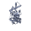
 Movie
Movie Controller


+ Open data

- Basic information
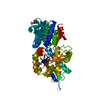
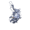
| Entry | Database: PDB / ID: 5w83 | |||||||||
|---|---|---|---|---|---|---|---|---|---|---|
| Title | Rpn8/Rpn11 dimer complex | |||||||||
 Components Components |
| |||||||||
 Keywords Keywords |  HYDROLASE / Protease HYDROLASE / Protease | |||||||||
| Function / homology |  Function and homology information Function and homology informationperoxisome fission / proteasome storage granule assembly / proteasome regulatory particle, lid subcomplex / mitochondrial fission / Cross-presentation of soluble exogenous antigens (endosomes) / TNFR2 non-canonical NF-kB pathway / Ub-specific processing proteases / proteasome binding / protein deubiquitination / proteasome storage granule ...peroxisome fission / proteasome storage granule assembly / proteasome regulatory particle, lid subcomplex / mitochondrial fission / Cross-presentation of soluble exogenous antigens (endosomes) / TNFR2 non-canonical NF-kB pathway / Ub-specific processing proteases / proteasome binding / protein deubiquitination / proteasome storage granule / proteasome assembly / Neutrophil degranulation / proteasome complex / metallopeptidase activity / ubiquitin-dependent protein catabolic process / proteasome-mediated ubiquitin-dependent protein catabolic process / ubiquitinyl hydrolase 1 / cysteine-type deubiquitinase activity / mitochondrion / metal ion binding / nucleus / cytosolSimilarity search - Function | |||||||||
| Biological species |  Saccharomyces cerevisiae (brewer's yeast) Saccharomyces cerevisiae (brewer's yeast) | |||||||||
| Method | X-RAY DIFFRACTION / SYNCHROTRON / MOLECULAR REPLACEMENT / Resolution: 1.554 Å | |||||||||
 Authors Authors | Dong, K.C. / Worden, E.J. / Martin, A. | |||||||||
| Funding support |  United States, 2items United States, 2items
| |||||||||
 Citation Citation | Journal: Mol. Cell / Year: 2017 Title: An AAA Motor-Driven Mechanical Switch in Rpn11 Controls Deubiquitination at the 26S Proteasome. Authors: Worden, E.J. / Dong, K.C. / Martin, A. | |||||||||
| History |
|
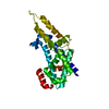
- Structure visualization
| Structure viewer | Molecule: MolmilJmol/JSmol |
|---|
- Downloads & links
-Download
| PDBx/mmCIF format | 5w83.cif.gz | 218.6 KB | Display | PDBx/mmCIF format |
|---|---|---|---|---|
| PDB format | pdb5w83.ent.gz | 174.6 KB | Display | PDB format |
| PDBx/mmJSON format | 5w83.json.gz | Tree view | PDBx/mmJSON format | |
| Others |  Other downloads Other downloads |
-Validation report
| Arichive directory | https://data.pdbj.org/pub/pdb/validation_reports/w8/5w83ftp://data.pdbj.org/pub/pdb/validation_reports/w8/5w83 | HTTPS FTP |
|---|
-Related structure data
| Related structure data | 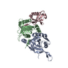 5u4pC 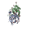 4o8xS C: citing same article ( S: Starting model for refinement |
|---|---|
| Similar structure data |


-Links
 PDBj
PDBj


- Assembly
| Deposited unit | 
| ||||||||
|---|---|---|---|---|---|---|---|---|---|
| 1 |
| ||||||||
| Unit cell |
|
-Components
| #1: Protein | Mass: 38365.508 Da / Num. of mol.: 1 Source method: isolated from a genetically manipulated source Source: (gene. exp.) Saccharomyces cerevisiae (strain ATCC 204508 / S288c) (yeast)Strain: ATCC 204508 / S288c / Gene: RPN8, YOR261C, O5360 / Production host:  Escherichia coli (E. coli) / References: UniProt: Q08723 Escherichia coli (E. coli) / References: UniProt: Q08723 |
|---|---|
| #2: Protein | Mass: 24880.621 Da / Num. of mol.: 1 Source method: isolated from a genetically manipulated source Source: (gene. exp.) Saccharomyces cerevisiae (strain ATCC 204508 / S288c) (yeast)Strain: ATCC 204508 / S288c / Gene: RPN11, MPR1, YFR004W / Production host: Escherichia coli (E. coli) / References: UniProt: P43588, ubiquitinyl hydrolase 1 |
| #3: Chemical | ChemComp-ZN /   Mass: 65.409 Da / Num. of mol.: 1 / Source method: isolated from a natural source / Formula: Zn Mass: 65.409 Da / Num. of mol.: 1 / Source method: isolated from a natural source / Formula: Zn |
| #4: Water | ChemComp-HOH / Water Mass: 18.015 Da / Num. of mol.: 207 / Source method: isolated from a natural source / Formula: H2O Mass: 18.015 Da / Num. of mol.: 207 / Source method: isolated from a natural source / Formula: H2O |
-Experimental details
-Experiment
| Experiment | Method: X-RAY DIFFRACTION / Number of used crystals: 1 |
|---|
- Sample preparation
| Crystal | Density Matthews: 1.44 Å3/Da / Density % sol: 14.31 % |
|---|---|
| Crystal grow | Temperature: 291 K / Method: vapor diffusion / Details: 1.5 M ammonium tartrate pH 6.8 |
-Data collection
| Diffraction | Mean temperature: 100 K |
|---|---|
| Diffraction source | Source: SYNCHROTRON / Site: ALS / Beamline: 8.3.1 / Wavelength: 1.11587 Å |
| Detector | Type: ADSC QUANTUM 315r / Detector: CCD / Date: Jun 4, 2015 |
| Radiation | Protocol: SINGLE WAVELENGTH / Monochromatic (M) / Laue (L): M / Scattering type: x-ray |
| Radiation wavelength | Wavelength: 1.11587 Å / Relative weight: 1 |
| Reflection | Resolution: 1.55→55.482 Å / Num. obs: 52965 / % possible obs: 99 % / Redundancy: 2 % / Net I/σ(I): 13.1 |
- Processing
| Software |
| |||||||||||||||||||||||||||||||||||||||||||||||||||||||||||||||||||||||||||||||||||||||||||||||||||||||||
|---|---|---|---|---|---|---|---|---|---|---|---|---|---|---|---|---|---|---|---|---|---|---|---|---|---|---|---|---|---|---|---|---|---|---|---|---|---|---|---|---|---|---|---|---|---|---|---|---|---|---|---|---|---|---|---|---|---|---|---|---|---|---|---|---|---|---|---|---|---|---|---|---|---|---|---|---|---|---|---|---|---|---|---|---|---|---|---|---|---|---|---|---|---|---|---|---|---|---|---|---|---|---|---|---|---|---|
| Refinement | Method to determine structure: MOLECULAR REPLACEMENT Starting model: 4O8X Resolution: 1.554→55.482 Å / SU ML: 0.24 / Cross valid method: FREE R-VALUE / σ(F): 1.33 / Phase error: 26.15 / Stereochemistry target values: ML
| |||||||||||||||||||||||||||||||||||||||||||||||||||||||||||||||||||||||||||||||||||||||||||||||||||||||||
| Solvent computation | Shrinkage radii: 0.9 Å / VDW probe radii: 1.11 Å / Solvent model: FLAT BULK SOLVENT MODEL | |||||||||||||||||||||||||||||||||||||||||||||||||||||||||||||||||||||||||||||||||||||||||||||||||||||||||
| Refinement step | Cycle: LAST / Resolution: 1.554→55.482 Å
| |||||||||||||||||||||||||||||||||||||||||||||||||||||||||||||||||||||||||||||||||||||||||||||||||||||||||
| Refine LS restraints |
| |||||||||||||||||||||||||||||||||||||||||||||||||||||||||||||||||||||||||||||||||||||||||||||||||||||||||
| LS refinement shell |
| |||||||||||||||||||||||||||||||||||||||||||||||||||||||||||||||||||||||||||||||||||||||||||||||||||||||||
| Refinement TLS params. | Method: refined / Origin x: 11.1005 Å / Origin y: 18.6694 Å / Origin z: 19.6081 Å
| |||||||||||||||||||||||||||||||||||||||||||||||||||||||||||||||||||||||||||||||||||||||||||||||||||||||||
| Refinement TLS group | Selection details: all |
